Annual Report 2022
Department of Radiological Technology
I. Radiological Diagnosis
Kanyu Ihara, Toshihiro Ishihara, Atsushi Urikura, Hirobumi, Nagasawa, Chieko Nagashima, Yoshiaki Miyazaki, Naoya Ikeno, Hiromi Suzuki, Jun Shishido, Makoto Mimatsu, Jun Torii, Yasutake Ishikawa, Minami Maruyama, Ryo Kikuchi, Hiroki Miyazaki, Eiko Taguchi, Yuya Kanai, Ryo Kawana, Yusuke Wakatsuki, Nao Ozaki, Ikuya Ozaki, Xiao Ang Tang, Seiya Mochizuki, Yuhei Shimizu, Yuta Miyamae, Akira Yoshida, Seiya Sato, Yuki Hirayama, Aya Shimoike, Chihiro Muto, Miku Kosaka, Ken Shimizu, Midori Nonaka, Kazuki Takaso, Satsuki Watanabe, Kazuki Matsubara, Yuki Tsunoda, Ai Arisue, Takumi Miyairi, Ayame Sugo, Yutaro Tsukahara, Tomoka Harada, Ryo Hosono, Yoshiki Ishihara, Sayaka Shimoda, Kaho Miyagi, Mio Kakinoki, Saya Muto, Banri Shinozaki, Madoka Hasegawa, Yuka Kondo, Yusuke Taira, Honoka Taira, Kodai Takahashi, Kotone Kumon, Kang SeokHyeon, Yoshitaka Murayama, Kei Terasaki
Introduction
Diagnostic radiology devices are essential for diagnosis, staging, response to treatment, and follow-up of cancer patients. The Department of Radiological Technology makes full use of the technology to provide precise medical images for patients. Human resource development of radiological technologists has always been, and continues to be, a theme in our department. We accept and educate residents in radiological technology. Further, we have accepted students and trainees of radiological technology from all over the world for global human resource development.
The Team and What We Do
The National Cancer Center Hospital has diagnostic imaging devices as shown in Table 1. We performed a total of 165,165 examinations as shown in Table 2 in FY2022.
The general radiography team established and defined fall and tripping hazard standards to prevent falls. We also evaluated the relationship between tube voltage and additive filters in general chest radiography and reported the results at an international conference.
The mammography team evaluated the retake rate and discussed positioning optimization.
The computed tomography (CT) team provided various additional information for diagnostic imaging using techniques such as deep learning-based reconstruction, iodine maps, and virtual monochromatic energy imaging. We have also researched and presented at international conferences on image quality and radiation dose in ultra-high-resolution CT and area detector CT.
The magnetic resonance imaging (MRI) team has prepared an operations and procedures manual to equalize the number of examination cases between each scanner and has also optimized the use of contrast media. In addition, we prepared the list for optimizing the acquisition protocols for clinical trials.
The interventional radiology team implemented close interprofessional information sharing to provide high-quality team care and has worked on creating and processing various 3-dimensional images for pre-interventional simulation to support diagnosis and treatment. We have begun to perform central venous port placement and removal procedures on an outpatient basis. In addition, novel applications were being developed in collaboration with some companies.
The gastrointestinal radiography team has engaged in comprehensive gastrointestinal examinations, performing contrast-enhanced gastrointestinal tract examinations and CT-colonography using X-TV and X-ray CT, respectively. The CT scanner was upgraded in May 2022, allowing further dose reductions in lung cancer screening. Further, a specialized team has been available for chest CT and chest X-ray examinations for COVID-19 patients.
The endoscopic radiography team worked to improve safety during procedures, improve the exam room environment, share information before exams, and run simulations in case of sudden changes through multidisciplinary collaboration.
The nuclear medicine team has completed the installation of a positron emission tomography (PET)/CT system, which allows for improved image quality and rapid acquisition. We also have filed legal proceedings for the renewal of a PET/CT system. A novel intravitreal radioisotope therapy for the treatment of unresectable pheochromocytoma and paraganglioma, called intravitreal nuclear medicine therapy for PPGL, has been initiated.
Table 1. Number of diagnostic imaging devices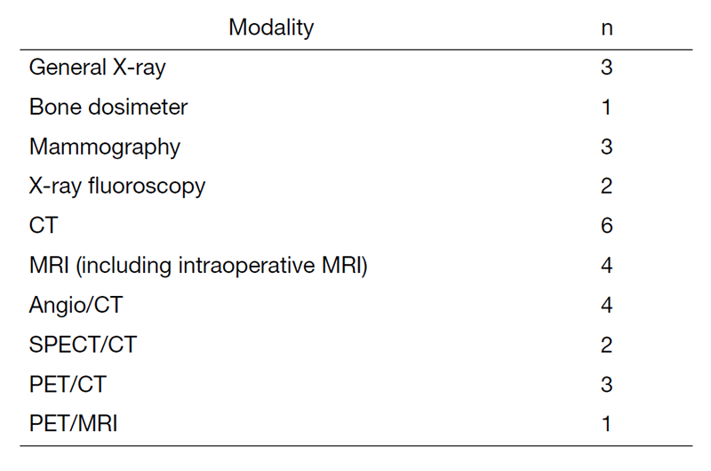
Table 2. Number of examination cases in FY2022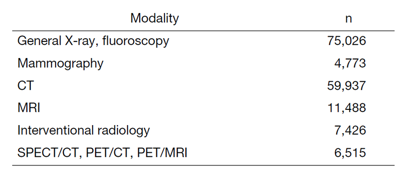
Research Activities
We conducted research in the fields of digital radiography (DR), CT, interventional radiology, and PET and presented the results at intra- and international conferences. We also conducted joint research with relevant companies as below.
Physical image data were analyzed for studies on the diagnosis of malignancies using dual-energy CT. We performed a phantom study to analyze the physical image characteristics of deep learning-based image processing software developed for reducing image noise in CT.
We also studied the use of dose management software to build a system that discloses and shares the medical exposure dose to/with patients.
Education
- Younger technologists participated in the training seminars hosted by the Ministry of Health, Labour and Welfare, the National Hospital Organization, and others; however, due to the COVID-19 pandemic, almost all of the seminars were conducted online.
- At the conference within the Department of Radiology Diagnostic Technology, senior technologists shared their specialized knowledge and contributed to improving skills.
- We have been conducting ongoing multi-site videoconferences with cancer hub hospitals in Japan. Even during the COVID-19 pandemic, this was effective for sharing information on radiological technology between institutes.
- We accepted hospital internship students from Juntendo University, the International University of Health and Welfare, Tokyo Metropolitan University, Kyorin University, Komazawa University, Tsukuba International University, and Yuanpei University of Medical Technology. Table 3 shows the results of accepted contracted training and practical training.
- We conducted personnel exchanges with Tokyo University Hospital and Kyoto University Hospital to provide mutually enriching education.
Table 3. Results of contracted training and practical training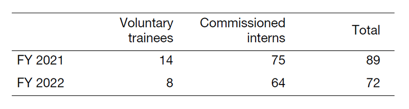
Future Prospects
The Department of Radiological Technology has established a residency program for radiological technologists beginning in FY 2022. We will provide continuing education to technologists specializing in cancer medicine.
II. Radiological Oncology
Kanyu Ihara, Tomonori Goka, Ako Aikawa, Toshimitsu Sofue, Miyuki Murata, Yoshihiro Shibata, Yusuke Watanabe, Tatsuya Sakasai, Junichi Kuwahara, Takayuki Mori, Yusuke Miyamoto, Mitsuhiro Kon, Masato Tanaka, Aya Shimoike, Yasunori Shuto, Hironori Murakami, Midori Nonaka, Masataka Ueda, Kaho Miyagi, Yusaku Kasai, Ruka Hata, Takuma Hanzawa, Koshin Arai, Masashi Kawamura, Homare Murata, Hiroko Sugaya
Introduction
The Radiological Oncology unit in Department of Radiological Technology is home to four linear accelerators, a CyberKnife, an MRI-guided radiotherapy system (MRIdian), image-guided radiation theraphy (IGRT), and image-guided brachytherapy (IGBT) systems, which we use to provide various radiotherapy treatments, including intensity modulated radiation therapy (IMRT), stereotactic radiation therapy (SRT), and high-dose-rate brachytherapy with IGBT.
The Team and What We Do
1) Two high-precision positioning systems (ExacTrac) enhanced the high-precision irradiation implementation system and improved treatment continuity by installing the same type of equipment side by side. The 2064 external radiation treatments in FY2022 consisted of 904 cases of VMAT and 432 cases of SRT; VMAT and SRT, which are high-precision radiation treatments, accounted for approximately 70% of the total.
2) The immediate adaptive radiotherapy system (MRIdian) was upgraded to a linac in July. Radiotherapy was administered to 112 patients, with a total of 349 online immediate adaptive radiotherapy (Adaptive Radiation Therapy) sessions.
3) A total of 164 cases were treated with stereotactic irradiation using a CyberKnife, consisting of 151 head-and-neck region and 13 trunk cases.
4) Total-body irradiation for bone marrow transplantation was performed in 54 cases.
5) Interstitial brachytherapy using a 106-Ru source and intertissued irradiation using a 198-Au source were performed in 12 and one cases, respectively.
6) The high-dose-rate intracavitary brachytherapy system was upgraded in August, making it possible to safely perform highly accurate treatment. Treatment using this system was performed in 132 patients with a total of 452 cases. The treatment planning CT system installed in the same room for IGBT was also upgraded to an 80-row LB CT system, reducing the acquisition time and improving the image quality.
7) Phase I clinical trials for boron neutron capture therapy (BNCT) were completed, and Phase II clinical trials were initiated.
8) To prevent COVID-19, we reinforced infection precautions and managed the business continuity plan.
Research Activities
We evaluated the properties of the MRIdian system. We also proposed a quality control method based on the establishment and implementation of quality control items required to maintain performance and long-term data.
Education
1) We had job rotations among the modalities corresponding to our educational programs.
2) We accepted hospital internship students from Juntendo University, the International University of Health and Welfare, Tokyo Metropolitan University, Kyorin University, Komazawa University, Tsukuba International University, and Yuanpei University of Medical Technology.
3) We have accepted students and trainees in radiological technology from all over the world for global human resource development.
4) One of the radiological technologists belonging to the radiotherapy team was concurrently appointed to the Section of Radiation Safety and Quality Assurance to learn about radiotherapy planning and quality control.
5) We reviewed and improved our workflow regarding the processes of radiotherapy.
6) We have launched a radiological technologist residency program to train and produce radiology technicians with highly specialized knowledge and skills. Two residents are hired to acquire the basic duties and the knowledge, skills, and attitude required of radiology technicians in a three-year program.
Future Prospects
1) Actively cooperate to achieve approval of the BNCT under the Pharmaceutical Affairs Law and early realization of its clinical use.
2) We will establish an efficient quality control system and education system to contribute to spreading the safe use of the MRIdian radiotherapy system.
3) We will aim for advanced radiotherapy with MRIdian system.
4) The system for RALS has been updated to a high dose rate system, and we will enable safe treatment and increase accurate results.
5) Devices in use for over 10 years will be replaced, and adaptive radiotherapy will be planned to provide stable and precise treatment.
6) We will continue to cooperate with the clinical trials conducted in the Department of Radiation Oncology.
List of papers published in 2022
Journal
1. Koretsune Y, Sone M, Sugawara S, Wakatsuki Y, Ishihara T, Hattori C, Fujisawa Y, Kusumoto M. Validation of a convolutional neural network for the automated creation of curved planar reconstruction images along the main pancreatic duct. Japanese journal of radiology, 41:228-234, 2023
2. Kon M, Okamoto H, Nakamura S, Iijima K, Chiba T, Takemori M, Nakayama H, Nakaichi T, Mikasa S, Fujii K, Urago Y, Ishikawa M, Sofue T, Katsuta S, Inaba K, Igaki H, Aso T. Planning study: prone versus supine position for stereotactic body radiotherapy in prostate by CyberKnife. Journal of radiation research, 64:186-194, 2023
3. Takemori M, Nakamura S, Sofue T, Ito M, Goka T, Miura Y, Iijima K, Chiba T, Nakayama H, Nakaichi T, Mikasa S, Takano Y, Kon M, Shuto Y, Urago Y, Nishitani M, Kashihara T, Takahashi K, Murakami N, Nishio T, Okamoto H, Chang W, Igaki H. Failure modes and effects analysis study for accelerator-based Boron Neutron Capture Therapy. Medical physics, 50:424-439, 2023
4. Nakaichi T, Okamoto H, Kon M, Takaso K, Aikawa A, Nakamura S, Ijima K, Chiba T, Nakayama H, Takemori M, Mikasa S, Fujii K, Urago Y, Goka T, Shimizu Y, Igaki H. Commissioning and performance evaluation of commercially available mobile imager for image guided total body irradiation. Journal of applied clinical medical physics, 24:e13865, 2023
5. Nakayama H, Okamoto H, Nakamura S, Iijima K, Chiba T, Takemori M, Nakaichi T, Mikasa S, Fujii K, Sakasai T, Kuwahara J, Miura Y, Fujiyama D, Tsunoda Y, Hanzawa T, Igaki H, Chang W. Film measurement and analytical approach for assessing treatment accuracy and latency in a magnetic resonance-guided radiotherapy system. Journal of applied clinical medical physics, 24:e13915, 2023
6. Arai T, Murata S, Watanabe Y, Ishihara T, Fukamizu Y, Takeda S, Ebata K, Watanabe Y, Takashima Y, Kaneko J. Fact-finding survey on the competencies and literacy of radiological technologists regarding radiation disasters. Journal of X-ray science and technology, 31:237-245, 2023
7. Urikura A, Miyauchi Y, Yoshida T, Ishita Y, Takiguchi K, Endo M, Aramaki T. Patient dose increase caused by posteroanterior CT localizer radiographs. Radiography (London, England : 1995), 29:334-339, 2023
8. Nishioka S, Okamoto H, Chiba T, Sakasai T, Okuma K, Kuwahara J, Fujiyama D, Nakamura S, Iijima K, Nakayama H, Takemori M, Tsunoda Y, Kaga K, Igaki H. Identifying risk characteristics using failure mode and effect analysis for risk management in online magnetic resonance-guided adaptive radiation therapy. Physics and imaging in radiation oncology, 23:1-7, 2022
9. Okamoto H, Igaki H, Chiba T, Shibuya K, Sakasai T, Jingu K, Inaba K, Kuroda K, Aoki S, Tatsumi D, Nakamura M, Kadoya N, Furuyama Y, Kumazaki Y, Tohyama N, Tsuneda M, Nishioka S, Itami J, Onishi H, Shigematsu N, Uno T. Practical guidelines of online MR-guided adaptive radiotherapy. Journal of radiation research, 63:730-740, 2022
10. Hasegawa A, Ishihara T, Pan T, Ropp AM, Winkler M, Sneider MB. Impact of pixel value truncation on image quality of low dose chest CT. Medical physics, 49:2979-2994, 2022
11. Ishita Y, Urikura A, Yoshida T, Takiguchi K, Ikegaya M. Inaccurate table height setting affects the organ-specific radiation dose in computed tomography. European journal of radiology, 151:110317, 2022
12. Miyazaki Y, Shimizu J, Kubo Y, Tabata N, Aso T. Quantitative classification of invasive and noninvasive breast cancer using dynamic magnetic resonance imaging of the mammary gland. Journal of clinical imaging science, 12:45, 2022
13. Urikura A, Yoshida T, Endo M, Asakura K, Sato R, Saiga A, Moriguchi M, Nakashima K, Aramaki T. Computed tomographic pulmonary angiography: Three cases of low-tube-voltage acquisition with a slow injection of contrast medium. Acta radiologica open, 11:20584601221131476, 2022