Home > Single-molecule imaging and super-resolution microscopy of tumor-derived extracellular vesicles (EVs)
Single-molecule imaging and super-resolution microscopy of tumor-derived extracellular vesicles (EVs)
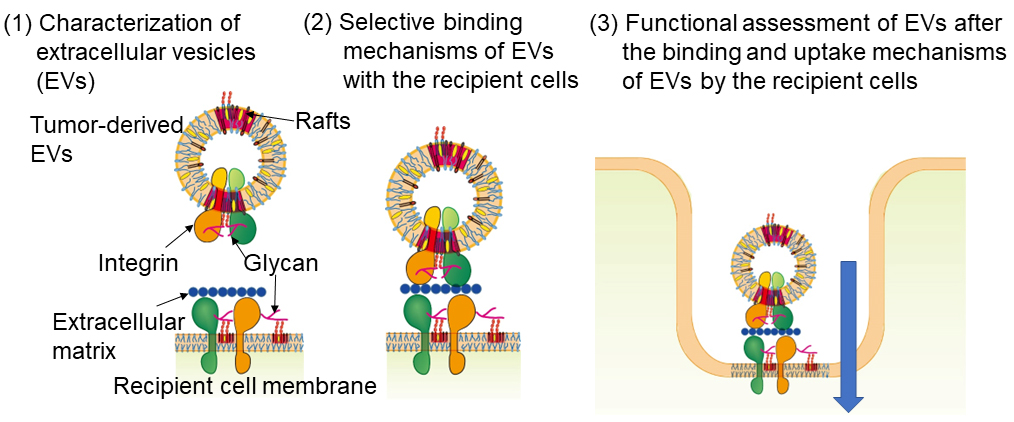
Recently, extracellular vesicles or EVs (40~200 nm in diameter) have attracted considerable attention as a carrier of intercellular communication. EVs are responsible for transporting various biological substances such as nucleic acids (mainly mRNA), proteins, lipids, and glycans between cells. Furthermore, it has been reported that after tumor-derived EVs are taken up by cells in other organs, an environment is formed around those cells that facilitates the metastasis of cancer cells. However, EVs are extremely heterogenous populations with a variety of subtypes, and methods that examine all EVs together cannot accurately study their properties. We fluorescently label EVs with marker molecules and observe the behaviors of individual EV particles (subtype by subtype). We are also attempting to elucidate the mechanism of interaction between EVs and molecules/structures on target cells by simultaneously observing various molecules and membrane structures on the target cell membrane and EVs with single-molecule imaging and super-resolution microscopy (see Figure).