Annual Report 2022
Division of Science and Technology for Endoscopy (Tsukiji Campus)
Shigetaka Yoshinaga, Yutaka Saito, Haruhisa Suzuki, Satoru Nonaka, Naoya Toyoshima, Yasuhiko Mizuguchi, Masayoshi Yamada, Susumu Hijioka, Nozomu Kobayashi, Yasuo Kakugawa, Seiichiro Abe, Hiroyuki Takamaru, Keiko Nakamura, Ichiro Oda, Taku Sakamoto
Introduction
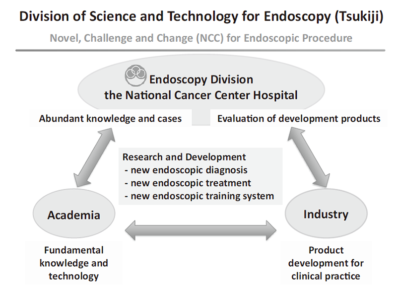
Since the birth of endoscope about half a century ago, endoscopes have undergone an era of fiber scopes. Now, with the introduction of videoscopes and high-definition systems, image quality has improved dramatically, and the target organs for observation and diagnosis have expanded from the stomach to include the esophagus, duodenum, colon, bronchus, biliary tract, and pharynx. The primary purpose of endoscopy is "observation and diagnosis" to detect lesions and check target organs; however, in recent years, "treatment" using endoscopes has become possible additionally. Nevertheless, currently, diagnosis is limited to focusing on morphological characteristics, and lesions that are missed occur at a certain rate, and difficult-to-treat lesions and treatment-related complications may also be encountered. In addition, the quality of endoscopic diagnosis and treatment often depends on the skill of endoscopists, and various issues remain to be resolved. To solve these issues and future demands, the Division of Science and Technology for Endoscopy, in collaboration with industry, government, and academia, is working to develop innovative endoscopic diagnosis, treatment, and training systems, and to disseminate them from Japan to the world (Figure 1).
Meanwhile, future demands include the development of innovative endoscopic diagnostic devices that visualize cancer characteristics, such as molecular imaging and functional imaging, and innovative endoscopic treatment techniques, such as endoscopic full-thickness resection. The Endoscopy Division of the National Cancer Center Hospital has a wealth of knowledge and clinical experience, which will play an effective role in the development of fundamental knowledge and technologies in academia, the development of products for actual clinical use and the evaluation and demonstration of developed products in industry.
The Team and What We Do
Activities began in fiscal year 2017, and in fiscal year 2022, each of the four teams (Diagnostic Team, Treatment Team, Training Team, and Endoscopic ultrasonography Team) will continue to develop innovative endoscopic diagnosis, treatment, and training systems, with research results being gradually obtained.
Diagnosis Team: Naoya Toyoshima, Yasuhiko Mizuguchi, Haruhisa Suzuki, Satoru Nonaka, Masayoshi Yamada, Taku Sakamoto
Treatment Team: Satoru Nonaka, Seiichiro Abe, Hiroyuki Takamaru
Training Team: Haruhisa Suzuki, Nozomu Kobayashi, Yasuo Kadokawa, Naoya Toyoshima, Hiroyuki Takamaru, Keiko Nakamura
Endoscopic Ultrasonography Team: Susumi Hijioka, Shigetaka Yoshinaga
Supervisors: Shigetaka Yoshinaga, Yutaka Saito
At the Olympus Lab in the Corporate Collaboration Lab of the National Cancer Center Research Institute, we collaborate with Olympus Corporation to develop new endoscopic diagnostic, therapeutic, and training devices. The details are currently confidential, but each team is working on 3 to 4 themes.
Research Activities
1) Diagnosis Team
Length measuring device: The shape of 69 lesions was measured, and a formula was developed to predict the depth of lesions based on indices such as tension, which was evaluated in 53 lesions for a prospective study. In addition, spectroscopic measurements of lesions were performed to evaluate the optical spectral slope and change in lesion height.
Bio-optical phantoms: Under a joint research agreement between Olympus Corporation and NCCH, we measured lesions in the esophagus, stomach, and large intestine and accumulated data.
RDI Pit Pattern Color Difference Analysis: We have demonstrated that the combination of Red Dichromatic Imaging (RDI) and indigo carmine spraying facilitates the recognition of colorectal tumor lesion pit patterns by color difference analysis, and have been working on the publication of the results.
Improvement of quality of examination: To improve the quality of intragastric examination, we created a prototype model of the stomach with LEDs attached to the current model and developed an application to evaluate it.
Prevention of contamination of the lens of endoscopes: We investigated the prevention of contamination of the field of view of endoscopes from three perspectives: "evaluation of cleanability", "search for degradation factors" and "examination of cleaning techniques". We used pseudo-fouling models for "evaluation of cleanability," and "search for degradation factors. For the "examination of cleaning techniques," we searched for lens coating agents from both water repellent and hydrophilic viewpoints.
2) Treatment team
Suturing Devices: In collaboration with Olympus Corporation, we developed a stapler for soft endoscopes, and we conducted chronic animal experiments in August to confirm its clinical efficacy. However, after a value study within Olympus Corporation, they reached the conclusion that it was not profitable due to cost and the number of cases, and development at Olympus Corporation was terminated.
Platform for colonoscopy: We have been exploring the needs and cost estimates for a "device that is attached to the anus and supports endoscopic procedures," as well as an image of the device to reduce costs.
Effectiveness verification of large-diameter forceps holes: A brainstorming session was held in October, and we are reviewing the promotion system within Olympus.
3) Training Team
Endoscopic Operability: We studied the usefulness of endoscopic technique training using an endoscopy simplified training simulator, and demonstrated that this simulator is useful in training-beginning physicians at the 103rd Annual Meeting of the Japanese Society of Gastrointestinal Endoscopy in May 2022. We then further explored new approaches using the endoscope simulator to simulate automatic operation by AI. Nevertheless, it is difficult for AI alone to complete the game autonomously, and we are now in the process of conducting a drastic review.
Colon insertion technology: In collaboration with Olympus, we are building a system to evaluate colonoscopy insertion techniques. As part of this review, we plan to present our efforts to quantify colonoscopy insertion operations using a training model at the 116th annual meeting of the Japan Gastroenterological Endoscopy Society, Kanto Chapter, in June 2023. In the future, as a new initiative, we plan to develop a new colonoscopy insertion training model based on the pain evaluation system developed by Tottori University and utilizing the know-how accumulated by Olympus and the three parties. We have conducted preliminary discussions with Tottori University.
Treatment Training Kit: For endoscopic treatment training, a pseudo gastrointestinal mucosa was created using konjak ingredients.
Education
Residents of the Endoscopy Division at NCCH participate in the meetings of each development team and in various development experiments to train and educate the next generation of people who will be responsible for endoscopic device development.
Future Prospects
In the Division of Science and Technology for Endoscopy, we would like to work on the development of innovative endoscopic diagnosis, treatment, and training systems in collaboration with industry, academia, and government to solve many current issues and future demands in daily clinical practice, and to disseminate them from Japan to the world.
List of papers published in 2022
Journal
1. Matsuzaki J, Kato K, Oono K, Tsuchiya N, Sudo K, Shimomura A, Tamura K, Shiino S, Kinoshita T, Daiko H, Wada T, Katai H, Ochiai H, Kanemitsu Y, Takamaru H, Abe S, Saito Y, Boku N, Kondo S, Ueno H, Okusaka T, Shimada K, Ohe Y, Asakura K, Yoshida Y, Watanabe SI, Asano N, Kawai A, Ohno M, Narita Y, Ishikawa M, Kato T, Fujimoto H, Niida S, Sakamoto H, Takizawa S, Akiba T, Okanohara D, Shiraishi K, Kohno T, Takeshita F, Nakagama H, Ota N, Ochiya T. Prediction of tissue-of-origin of early stage cancers using serum miRNomes. JNCI cancer spectrum, 7:pkac080, 2023
2. Suzuki H, Ono H, Hirasawa T, Takeuchi Y, Ishido K, Hoteya S, Yano T, Tanaka S, Toya Y, Nakagawa M, Toyonaga T, Takemura K, Hirasawa K, Matsuda M, Yamamoto H, Tsuji Y, Hashimoto S, Yuki M, Oyama T, Takenaka R, Yamamoto Y, Naito Y, Yamamoto K, Kobayashi N, Kawahara Y, Hirano M, Koizumi S, Hori S, Tajika M, Hikichi T, Yao K, Yokoi C, Ohnita K, Hisanaga Y, Sumiyoshi T, Kitamura S, Tanaka H, Shimoda R, Shimazu T, Takizawa K, Tanabe S, Kondo H, Iishi H, Ninomiya M, Oda I. Long-term Survival After Endoscopic Resection For Gastric Cancer: Real-world Evidence From a Multicenter Prospective Cohort. Clinical gastroenterology and hepatology, 21:307-318.e2, 2023
3. Fukushi G, Yamada M, Kakugawa Y, Gotoh M, Tanabe N, Ushiama M, Watanabe T, Yamazaki T, Matsumoto M, Hirata M, Nakajima T, Sugano K, Yoshida T, Matsuda T, Igarashi Y, Saito Y. Genotype-phenotype correlation of small-intestinal polyps on small-bowel capsule endoscopy in familial adenomatous polyposis. Gastrointestinal endoscopy, 97:59-68.e7, 2023
4. Hiki K, Ogata D, Hisada I, Sakamoto T, Hiroyuki T, Yamakawa K, Namikawa K, Takahashi A, Saito Y, Yamazaki N. Minimally invasive surgery combined with excision and endoscopic submucosal dissection for anorectal melanoma. The Journal of dermatology, 50:e26-e27, 2023
5. Hirata S, Toyoshima N, Takamaru H, Yamada M, Kobayashi N, Kozu T, Saito Y. Underwater endoscopic mucosal resection with submucosal injection. Endoscopy, 55:E70-E71, 2023
6. Takamaru H, Saito Y, Toyoshima N, Yamada M, Sakamoto T, Matsuda T. Polyglycolic acid sheet with clipping for closing delayed perforation after colonic endoscopic submucosal dissection. Endoscopy, 55:E211-E213, 2023
7. Uraoka T, Uedo N, Oyama T, Saito Y, Yahagi N, Fujimoto A, Kawahara Y, Mabe K, Hikichi T, Yamamoto Y, Tajiri H. Efficacy and Safety of a Novel Hemostatic Peptide Solution During Endoscopic Submucosal Dissection: A Multicenter Randomized Controlled Trial. The American journal of gastroenterology, 118:276-283, 2023
8. Saito Y. Pathologic sm2 carries a moderate risk of metastases even without other unfavorable factors, but positive horizontal margins have low local recurrence risk after en bloc resection. Endoscopy, 55:252-254, 2023
9. Kadota T, Ishihara R, Hatta W, Yoshida M, Kanzaki H, Kikuchi D, Ono Y, Abe S, Yamamoto Y, Yoshio T, Urabe Y, Yamaguchi N, Nagami Y, Iizuka T, Takahashi H, Oyama T, Yano T. Multi-institutional questionnaire on treatment strategies for superficial entire circumferential esophageal squamous cell carcinoma. DEN open, 3:e206, 2023
10. Hirai Y, Abe S, Makiguchi ME, Sekiguchi M, Nonaka S, Suzuki H, Yoshinaga S, Saito Y. Endoscopic Resection of Undifferentiated Early Gastric Cancer. Journal of gastric cancer, 23:146-158, 2023
11. Yamada M, Shino R, Kondo H, Yamada S, Takamaru H, Sakamoto T, Bhandari P, Imaoka H, Kuchiba A, Shibata T, Saito Y, Hamamoto R. Robust automated prediction of the revised Vienna Classification in colonoscopy using deep learning: development and initial external validation. Journal of gastroenterology, 57:879-889, 2022
12. Kakeji Y, Ishikawa T, Suzuki S, Akazawa K, Irino T, Miyashiro I, Ono H, Suzuki H, Tanabe S, Kadowaki S, Muro K, Fukagawa T, Nunobe S, Wada T, Katai H, Kodera Y. A retrospective 5-year survival analysis of surgically resected gastric cancer cases from the Japanese Gastric Cancer Association nationwide registry (2001-2013). Gastric cancer, 25:1082-1093, 2022
13. Shiroma H, Shiba S, Erawijantari PP, Takamaru H, Yamada M, Sakamoto T, Kanemitsu Y, Mizutani S, Soga T, Saito Y, Shibata T, Fukuda S, Yachida S, Yamada T. Surgical Treatment for Colorectal Cancer Partially Restores Gut Microbiome and Metabolome Traits. mSystems, 7:e0001822, 2022
14. Kasuga K, Oda I, Nonaka S, Abe S, Suzuki H, Uraoka T, Saito Y. Endoscopic complete closure of duodenal mucosal defects using a clip with a looped thread after endoscopic resection. Endoscopy, 54:E135-E136, 2022
15. Kasuga K, Saito Y, Takamaru H, Yamada M, Sakamoto T, Sekine S, Uraoka T. Optical real-time biopsy by endocytoscopy: a case of sessile serrated lesion with dysplasia. Endoscopy, 54:E249-E251, 2022
16. Kasuga K, Abe S, Oda I, Yoshinaga S, Suzuki H, Uraoka T, Saito Y. Guidewire-assisted technique for gastroscope insertion through stricture of Zenker’s diverticulum for esophageal endoscopic submucosal dissection. Endoscopy, 54:E279-E280, 2022
17. Sakamoto T, Nakashima H, Nakamura K, Nagahama R, Saito Y. Performance of Computer-Aided Detection and Diagnosis of Colorectal Polyps Compares to That of Experienced Endoscopists. Digestive diseases and sciences, 67:3976-3983, 2022
18. Harai S, Hijioka S, Maruki Y, Ohba A, Nagashio Y, Okusaka T, Saito Y. Endoscopic ultrasound-guided hepaticoduodenostomy with anterograde stenting for recurrent hepatic hilar obstruction. Endoscopy, 54:E398-E400, 2022
19. Abe S, Makiguchi ME, Nonaka S, Suzuki H, Yoshinaga S, Saito Y. Emerging texture and color enhancement imaging in early gastric cancer. Digestive endoscopy, 34:714-720, 2022
20. Koyama Y, Fukuzawa M, Kono S, Madarame A, Morise T, Uchida K, Yamaguchi H, Sugimoto A, Nagata N, Kawai T, Takamaru H, Sekiguchi M, Yamada M, Sakamoto T, Matsuda T, Saito Y, Itoi T. Diagnostic efficacy of the Japan NBI Expert Team classification with dual-focus magnification for colorectal tumors. Surgical endoscopy, 36:5032-5040, 2022
21. Kobayashi N, Takeuchi Y, Ohata K, Igarashi M, Yamada M, Kodashima S, Hotta K, Harada K, Ikematsu H, Uraoka T, Sakamoto N, Doyama H, Abe T, Katagiri A, Hori S, Michida T, Yamaguchi T, Fukuzawa M, Kiriyama S, Fukase K, Murakami Y, Ishikawa H, Saito Y. Outcomes of endoscopic submucosal dissection for colorectal neoplasms: Prospective, multicenter, cohort trial. Digestive endoscopy, 34:1042-1051, 2022
22. Takamaru H, Saito Y, Hammoud GM, Mizuguchi Y, Cho H, Sekiguchi M, Yamada M, Sakamoto T, Matsuda T. Comparison of postpolypectomy bleeding events between cold snare polypectomy and hot snare polypectomy for small colorectal lesions: a large-scale propensity score-matched analysis. Gastrointestinal endoscopy, 95:982-989.e6, 2022
23. Uraoka T, Takizawa K, Tanaka S, Kashida H, Saito Y, Yahagi N, Yamano HO, Saito S, Hisabe T, Yao T, Watanabe M, Yoshida M, Saitoh Y, Tsuruta O, Igarashi M, Toyonaga T, Ajioka Y, Fujimoto K, Inoue H. Guidelines for Colorectal Cold Polypectomy (supplement to "Guidelines for Colorectal Endoscopic Submucosal Dissection/Endoscopic Mucosal Resection"). Digestive endoscopy, 34:668-675, 2022
24. Kitamura H, Hijioka S, Nagashio Y, Ban D, Esaki M, Okusaka T, Saito Y. A case of high grade pancreatic intraepithelial neoplasia diagnosed by endoscopic ultrasound-guided fine needle aspiration. Endoscopy, 54:E628-E630, 2022
25. Abe S, Tomizawa Y, Saito Y. Can artificial intelligence be your angel to diagnose early gastric cancer in real clinical practice? Gastrointestinal endoscopy, 95:679-681, 2022
26. Abe S, Hirai Y, Uozumi T, Makiguchi ME, Nonaka S, Suzuki H, Yoshinaga S, Oda I, Saito Y. Endoscopic resection of esophageal squamous cell carcinoma: Current indications and treatment outcomes. DEN open, 2:e45, 2022
27. Abe S, Yamazaki T, Hisada IT, Makiguchi ME, Yoshinaga S, Sato T, Nonaka S, Suzuki H, Oda I, Saito Y. Visibility of early gastric cancer in texture and color enhancement imaging. DEN open, 2:e46, 2022
28. Saito Y, Ono A, García VAJ, Mizuguchi Y, Hisada I, Takamaru H, Yamada M, Sekiguchi M, Makiguchi M, Sekine S, Abe S. Diagnosis and treatment of colorectal tumors: Differences between Japan and the West and future prospects. DEN open, 2:e66, 2022
29. Koga T, Hijioka S, Nagashio Y, Ohba A, Maruki Y, Yoshinari M, Hisada Y, Harai S, Kitamura H, Maehara K, Murashima Y, Kawasaki Y, Kawahara S, Takeshita K, Yamada N, Satake T, Kondo S, Morizane C, Ueno H, Okusaka T, Saito Y. Endoscopic ultrasound-guided choledochoduodenostomy without fistula dilation using a stent with a 5.9-Fr delivery system: Comparison to a conventional procedure with fistula dilation. DEN open, 2:e56, 2022
30. Ma X, Kawashima K, Saito Y. Using the string-clip method to retrieve the resected specimen allowed a clear observation of the colon and detection of a new lesion. Digestive endoscopy, 34:e77-e78, 2022
31. Abe S, Sekiguchi M. It is time to tailor endoscopic resection for early gastric cancer: Evaluate not only lesion but also patient. Digestive endoscopy, 34:826-827, 2022
32. Kawasaki Y, Hijioka S, Saito Y. Modified double-guidewire technique using a new double-lumen catheter and 0.018-inch guidewire for difficult biliary cannulation. Digestive endoscopy, 34:e71-e72, 2022
33. Saito Y, Yamada M, Mori Y. Although depth prediction of colorectal cancer with artificial intelligence is clinically relevant, standardization of histopathologic diagnosis should also be taken care of. Gastrointestinal endoscopy, 95:1195-1197, 2022
34. Toyoshima N, Abe S, Saito Y. In addition to free deep margins, R0 resection should be required for T1 colorectal cancers to inform further surgical resection. Endoscopy international open, 10:E291-E292, 2022
35. Yachida T, Matsuda T, Sakamoto T, Nakajima T, Kakugawa Y, Maeshima AM, Taniguchi H, Kushima R, Tobinai K, Kobara H, Masugata H, Masaki T, Saito Y. Endoscopic features of colorectal lymphoma according to histological type. JGH open, 6:257-262, 2022
36. Kawasaki Y, Hijioka S, Maehara K, Tamada K, Okusaka T, Saito Y. Endoscopic ultrasound-guided intra-afferent loop entero-enterostomy using a forward-viewing echoendoscope and insertion of a metal stent. Endoscopy, 54:E815-E817, 2022
37. Sakamoto T, Akiyama S, Narasaka T, Suzuki H, Sekine S, Saito Y, Tsuchiya K. Anal Intraepithelial Neoplasia: Precursor of Anal Squamous Cell Carcinoma. Journal of the anus, rectum and colon, 6:92-99, 2022
38. Ainechi D, Misawa M, Barua I, Larsen SLV, Paulsen V, Garborg KK, Aabakken L, Tønnesen CJ, Løberg M, Kalager M, Kudo SE, Hotta K, Ohtsuka K, Saito S, Ikematsu H, Saito Y, Matsuda T, Itoh H, Mori K, Bretthauer M, Mori Y. Impact of artificial intelligence on colorectal polyp detection for early-career endoscopists: an international comparative study. Scandinavian journal of gastroenterology, 57:1272-1277, 2022
39. Kuriki Y, Yoshioka T, Kamiya M, Komatsu T, Takamaru H, Fujita K, Iwaki H, Nanjo A, Akagi Y, Takeshita K, Hino H, Hino R, Kojima R, Ueno T, Hanaoka K, Abe S, Saito Y, Nakajima J, Urano Y. Development of a fluorescent probe library enabling efficient screening of tumour-imaging probes based on discovery of biomarker enzymatic activities. Chemical science, 13:4474-4481, 2022
40. Saito Y, Takamaru H, Toyoshima N. Resection depth: a very important advantage for underwater EMR. Endoscopy international open, 10:E729-E730, 2022
41. Ohata K, Kobayashi N, Sakai E, Takeuchi Y, Chino A, Takamaru H, Kodashima S, Hotta K, Harada K, Ikematsu H, Uraoka T, Murakami T, Tsuji S, Abe T, Katagiri A, Hori S, Michida T, Suzuki T, Fukuzawa M, Kiriyama S, Fukase K, Murakami Y, Ishikawa H, Saito Y. Long-term Outcomes After Endoscopic Submucosal Dissection for Large Colorectal Epithelial Neoplasms: A Prospective, Multicenter, Cohort Trial From Japan. Gastroenterology, 163:1423-1434.e2, 2022
42. Hoshino Y, Hanaoka K, Sakamoto K, Yasunaga M, Kojima T, Kotani D, Nomoto A, Sasaki E, Komatsu T, Ueno T, Takamaru H, Saito Y, Seto Y, Urano Y. Molecular design of near-infrared (NIR) fluorescent probes targeting exopeptidase and application for detection of dipeptidyl peptidase 4 (DPP-4) activity. RSC chemical biology, 3:859-867, 2022
43. Yamamoto Y, Yoshida N, Yano T, Horimatsu T, Uedo N, Kawata N, Kanzaki H, Hori S, Yao K, Abe S, Katada C, Yokoi C, Ohata K, Doyama H, Yoshimura K, Ishikawa H, Muto M. Assessment of Outcomes From 1-Year Surveillance After Detection of Early Gastric Cancer Among Patients at High Risk in Japan. JAMA network open, 5:e2227667, 2022
44. Odagiri H, Hatta W, Tsuji Y, Yoshio T, Yabuuchi Y, Kikuchi D, Tsuji S, Nagami Y, Hikichi T, Kobayashi M, Morita Y, Sumiyoshi T, Iguchi M, Tomida H, Inoue T, Mikami T, Hasatani K, Nishikawa J, Matsumura T, Nebiki H, Nakamatsu D, Ohnita K, Suzuki H, Ueyama H, Hayashi Y, Sugimoto M, Yamaguchi S, Michida T, Yada T, Asahina Y, Narasaka T, Kuribayashi S, Kiyotoki S, Mabe K, Fujishiro M, Masamune A, Hoteya S. Bleeding following Endoscopic Submucosal Dissection for Early Gastric Cancer in Surgically Altered Stomach. Digestion, 103:428-437, 2022
45. Messmann H, Bisschops R, Antonelli G, Libânio D, Sinonquel P, Abdelrahim M, Ahmad OF, Areia M, Bergman JJGHM, Bhandari P, Boskoski I, Dekker E, Domagk D, Ebigbo A, Eelbode T, Eliakim R, Häfner M, Haidry RJ, Jover R, Kaminski MF, Kuvaev R, Mori Y, Palazzo M, Repici A, Rondonotti E, Rutter MD, Saito Y, Sharma P, Spada C, Spadaccini M, Veitch A, Gralnek IM, Hassan C, Dinis-Ribeiro M. Expected value of artificial intelligence in gastrointestinal endoscopy: European Society of Gastrointestinal Endoscopy (ESGE) Position Statement. Endoscopy, 54:1211-1231, 2022
46. Kahaleh M, Bhagat V, Dellatore P, Tyberg A, Sarkar A, Shahid HM, Andalib I, Alkhiari R, Gaidhane M, Kedia P, Nieto J, Kumta NA, Dixon RE, Salameh H, Mavrogenis G, Bassioukas S, Abe S, Arentes VN, Morita FH, Sakai P, de Moura EG. Subepithelial tumors: How does endoscopic full-thickness resection & submucosal tunneling with endoscopic resection compare with laparoscopic endoscopic cooperative surgery? Endoscopy international open, 10:E1491-E1496, 2022