Annual Report 2022
Department of Diagnostic Radiology
Tatsushi Kobayashi, Masayuki Yamaguchi, Hirofumi Kuno, Yasunori Arai, Kaoru Shimada, Tomoaki Sasaki, Takashi Hiyama, Shioto Oda, Takahiro Morita, Yohei Takei, Yusuke Miyasaka, Toshihiro Horii, Akihito Nakajima.
Introduction
The Department of Diagnostic Radiology is committed to advancing medical science through a dual emphasis on innovative, image-guided patient care and pioneering research. The department conducts approximately 140,000 inpatient and outpatient diagnostic imaging studies each year and is actively engaged in both clinical and translational research efforts. This report provides an overview of the department's facilities, workforce, research activities, and contributions to clinical trials.
The Team and What We Do
Our department is equipped with state-of-the-art imaging technologies, including four multi-slice CT scanners—one of which is an ultra-high-resolution CT—two area detector CT scanners, one dual-source CT, two 3T MRI systems, and one interventional radiology (IR) CT system. We also operate a multi-axis c-arm CT system, two gamma cameras with single photon emission CT (SPECT) capabilities, two digital radiographic (DR) systems for fluoroscopy, two mammography (MMG) units, and four computed radiographic (CR) systems. Our IR-CT systems leverage digital subtraction angiography in conjunction with multi-detector computerized tomography (MDCT), with one system featuring a 320 multi-slice CT scanner. The addition of a positron emission tomography (PET) scanner and a baby cyclotron has facilitated tumor imaging via 18F-FDG (fluorodeoxyglucose). These digital platforms enhance the efficiency and effectiveness of routine diagnostic procedures.
This department consists of ten staff radiologists, a senior resident and a resident. As part of our routine activities, every effort is made to produce an integrated report covering almost all examinations, such as MMG, contrast radiological procedures, CT, MRI, RI, PET, angiography and IR, primarily transarterial chemoembolization (TACE). Statistical data for the number of cases examined in 2022 can be found in Tables 1 and 2. Several conferences are routinely held at our department including pre- and postoperative conferences. Our department also contributes to deciding the treatment strategy through image presentations at the weekly tumor board conference.
Table 1. Number of examinations in 2022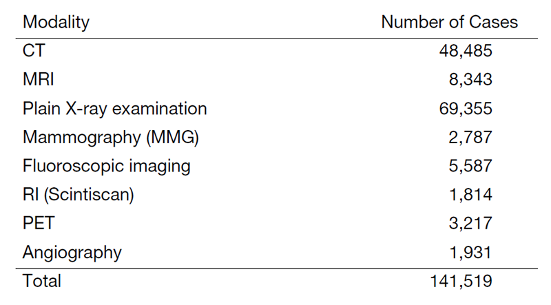
Table 2. Number of Interventional Radiology Procedures in 2022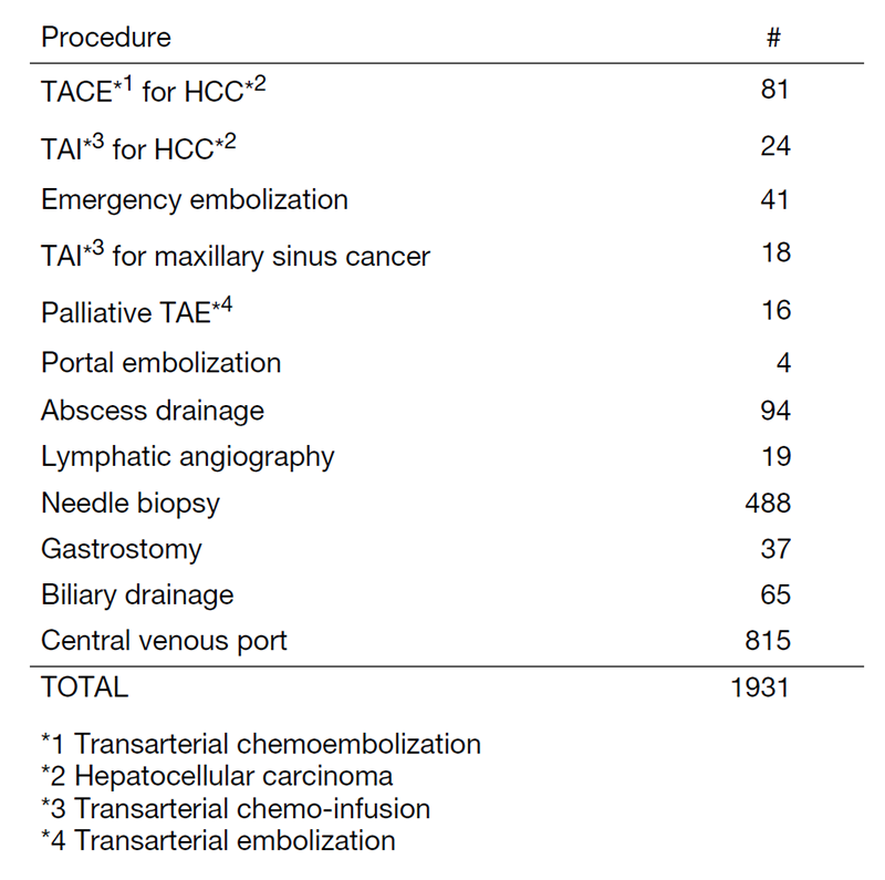
Research Activities
Imaging Technologies:
Our research primarily targets the development and clinical validation of next-generation imaging technologies. Special attention is given to Ultra-High-Resolution CT (UHR CT), Dual-Energy CT (DECT), and Area-Detector CT (ADCT), particularly for oncological applications. We are also exploring the utility of photon-counting CT technologies that provide both high spatial resolution and superior signal-to-noise ratios, thus enabling low-dose radiation protocols without compromising diagnostic accuracy. Furthermore, these technologies enable precise material differentiation through spectral imaging, thus augmenting our diagnostic confidence in cancer assessment.
Artificial Intelligence and Radiomics:
In addition to hardware innovations, our research also extends to the software domain, where we are exploring the incorporation of artificial intelligence (AI) algorithms and radiomics in medical imaging. Radiomics allows for a quantitative analysis of image features, offering deeper insights into disease processes beyond what is visually discernible. We are engaged in research projects that aim to correlate tumor-specific radiomic attributes with clinical endpoints such as treatment efficacy. Active partnerships with the industry are in progress to drive the development of AI algorithms for diagnostic imaging.
Clinical Trials
We are conducting a multicenter clinical trial, "The single-armed confirmatory trial for immediate effectivity and safety of palliative arterial embolization for painful bone metastases. (JIVROSG/J-SUPPORT 1903)", as a representative facility, which was started in March 2021. The purpose of this trial is to verify the safety and immediate effect of transarterial embolization as a palliative treatment for painful bone metastases and to establish it as a standard treatment. This research is funded by the Japan Agency for Medical Research and Development (AMED).
List of papers published in 2022
Journal
1. Sakai M, Nishimura B, Hiyama T, Kuno H, Shinozaki T, Sakamoto N, Nakajima T. Imaging of diffuse fibroepithelial polyps on surgical free flap in oral cancer patients: two case reports. Neuroradiology, 65:815-818, 2023
2. Funasaka C, Naito Y, Kusuhara S, Nakao T, Nakajima H, Kawamoto M, Baba K, Mamishin K, Kondoh C, Harano K, Matsubara N, Hosono A, Sasaki T, Kawasaki T, Mukohara T. Clinical features of CDK4/6 inhibitor-related interstitial lung disease in patients with breast cancer: a case series study. Japanese journal of clinical oncology, 53:105-114, 2023
3. Yamaguchi M, Kojo K, Akatsuka M, Haishi T, Kobayashi T, Nakajima T, Nishiyama H, Fujii H. High Resolution MR Imaging of the Testis Using a Small Radiofrequency Coil. Magnetic resonance in medical sciences, 22:127-136, 2023
4. Nakama R, Arai Y, Horii T, Kobayashi T. Air Embolism with Electrocardiogram Abnormality after Lung Biopsy. Internal medicine (Tokyo, Japan), 62:1383-1384, 2023
5. Masuoka S, Hiyama T, Kuno H, Sekiya K, Sakashita S, Kobayashi T. Imaging Approach for Cervical Lymph Node Metastases from Unknown Primary Tumor. Radiographics, 43:e220071, 2023
6. Ikeda M, Arai Y, Inaba Y, Tanaka T, Sugawara S, Kodama Y, Aramaki T, Anai H, Morita S, Tsukahara Y, Seki H, Sato M, Kamimura K, Azama K, Tsurusaki M, Sugihara E, Miyazaki M, Kobayashi T, Sone M. Conventional or Drug-Eluting Beads? Randomized Controlled Study of Chemoembolization for Hepatocellular Carcinoma: JIVROSG-1302. Liver cancer, 11:440-450, 2022
7. Takahashi S, Ohno I, Ikeda M, Konishi M, Kobayashi T, Akimoto T, Kojima M, Morinaga S, Toyama H, Shimizu Y, Miyamoto A, Tomikawa M, Takakura N, Takayama W, Hirano S, Otsubo T, Nagino M, Kimura W, Sugimachi K, Uesaka K. Neoadjuvant S-1 With Concurrent Radiotherapy Followed by Surgery for Borderline Resectable Pancreatic Cancer: A Phase II Open-label Multicenter Prospective Trial (JASPAC05). Annals of surgery, 276:e510-e517, 2022
8. Kudo M, Gotohda N, Sugimoto M, Konishi M, Takahashi S, Kobayashi S, Kobayashi T. The Assessment of Regional Liver Function Before Major Hepatectomy Using Magnetic Resonance Imaging. The American surgeon, 88:2353-2360, 2022
9. Nakama R, Arai Y, Hosoi K, Kobayashi T. Computed Tomography-Guided Percutaneous Intranodal Mesenteric Lymphangiography for the Identification of a Lymphatic Leak. Journal of vascular and interventional radiology, 33:854-855, 2022
10. Nakama R, Arai Y, Kobayashi T. Sedation With Dexmedetomidine During Lymphangiography and Thoracic Duct Embolization in an Elderly Man. Cureus, 14:e24466, 2022
11. Kaito D, Yamamoto R, Nakama R, Hashizume K, Ueno K, Sasaki J. D-dimer for screening of aortic dissection in patients with ST-elevation myocardial infarction. The American journal of emergency medicine, 59:146-151, 2022
12. Kujirai D, Fujii R, Kaito D, Nakama R, Izawa Y. Blast Injuries by an Improvised Explosive Device in Japan: A Case Report. Cureus, 14:e32118, 2022