Annual Report 2022
Department of Diagnostic Radiology
Masahiko Kusumoto, Miyuki Sone, Gen Iinuma, Nachiko Uchiyama, Hirokazu Watanabe, Mototaka Miyake, Shunsuke Sugawara, Kimiteru Ito, Yuko Kubo, Chihiro Ito, Miyako Morooka, Sachiko Nakano, Nao Kikkawa, Shintaro Kimura, Sawako Kaku, Takumi Oshima, Mizuki Ozawa, Tomoya Tanishima
Introduction
The Department of Diagnostic Radiology provides a wide range of modalities, including interventional radiology (IR), general radiology, computed tomography (CT), magnetic resonance imaging (MRI), mammography, and nuclear medicine imaging (Table 1).
In 2014, we launched the Interventional Radiology Center to facilitate the widespread proliferation of IR in Japan and to provide various IR treatments for patients referred from other hospitals or clinics.
We seek individuals with outstanding leadership capabilities, proven academic and administrative experience, a vision to build and sustain programs at the forefront of imaging research, and a commitment to clinical experience.
Table 1. Number of Examination per Modality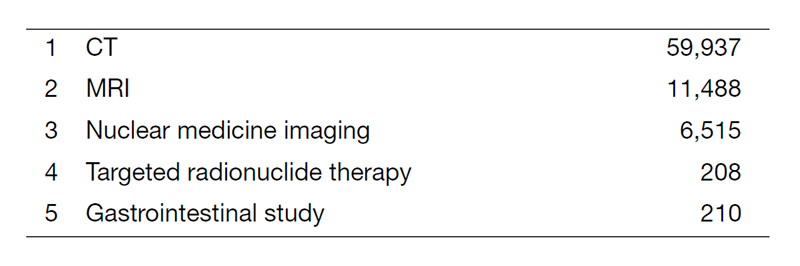
Table 2. Type of Percutaneous Interventional Radiology Procedure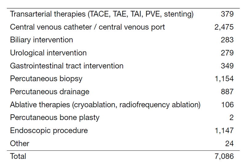
The Team and What We Do
In 2022, a total of 16 board-certified radiologists and two physicians performed routine radiological diagnosis, including IR procedures. We provided a total of 78,358 radiological diagnoses, the details of which are shown in Table 1. Furthermore, we implemented artificial intelligence software for diagnostic support in interpreting radiological images. A total of 7,086 IR procedures were performed from April 2022 to March 2023 at the center (Table 2). Furthermore, we had several consultation phone calls regarding the indications for various IR procedures from all over the country.
Research Activities
The CT colonography (CTC) has been successfully introduced as an effective option for preoperative staging and colorectal screening in our center. Approximately 700 patients and/or candidates were examined in 2022. We are developing artificial intelligence software for colorectal tumors, including flat lesions. The primary purpose of our CTC research work is to conduct a multicenter trial to establish evidence regarding fully digitalized CTC for a colorectal screening system in Japan.
A multi-tracer consisting of F-18 FDG, and Cu-64 ATSM PET/CT imaging has been investigated for the treatment of patients with cancer to improve the sensitivity and specificity of detecting tumor sites or tumor characteristics. F-18 FDG PET allows us to calculate the glucose metabolic rate of the tumor site and to predict the treatment response in patients treated with immune checkpoint inhibitors. Cu-64 ATSM PET/CT has been conducted in over 16 patients with brain tumor through the clinical research. In addition, PET/MRI has been applied to more than 1000 cases of cancer patients per year and proved itself to be a powerful tool for managing malignancy. For cancer treatment using radioisotope, internal radiotherapy was carried out using radioactive iodine (I-131) chloride, Zevalin, Lutathera, Xofigo, and Raiatt.
In the "Public/Private R&D Investment Strategic Expansion PrograM (PRISM)" project for the development of artificial intelligence to accelerate the creation of new drugs, we provided 2,000 chest CT images and diagnostic reports to build an integrated database for lung cancer treatment. We have also developed an annotation tool for radiological images to efficiently develop deep learning models.
We have explored the efficacy of ultra-high-resolution computed tomography (U-HRCT), which had been developed in the NCC. The research topics related to U-HRCT include: staging and evaluating the treatment efficacy of head and neck cancers; diagnostic accuracy of invasion to the pancreatic neural plexus in patients with pancreatic cancer, and simulation of IR procedures using the U-HRCT data. In addition, we have started developing radiomics and artificial intelligence for the detection and characterization of pancreatic tumor.
A major research theme in IR is the establishment of an evidence base for IR. We have conducted multi-institutional clinical trials as a steering organization as mentioned hereunder. The in-house research topics include: a preliminary report of PTBD in prone position for potential collaboration with endoscopic intervention, the necessity of the anticoagulation therapy after IVC stent placement in cancer patients, central venous ports in infants: review of a 11-year experience, analysis of risk factors for major bleeding complications after percutaneous liver biopsy, feasibility of TAE prior to renal cryoablation in RCC patients with chronic renal failure, and weight of specimens obtained with seven different biopsy needles. We have also developed novel IR techniques and software including palliative stent placement for patients with multiple bowel obstructions, percutaneous peritoneal lavage for patients with pancreatic cancer before pancreatectomy, and a navigation system for vascular embolization and tumor ablation.
Clinical Trials
We have led a multi-institutional cooperative study group of interventional radiology (JIVROSG: Japan Interventional Radiology in Oncology Study Group) since 2002 as a steering organization of 107 participating domestic institutions. A total of 29 studies were completed, and 22 were published. Currently, three clinical trials are ongoing.
In the nuclear medicine field, one clinical trial is ongoing: a phase I study evaluating the safety of Cu-64 ATSM for brain tumors. We also started developing another phase I study protocol of a new clinical trial evaluating the safety of Cu64 NCAB PET/CT for pancreatic cancer.
Education
The clinical education and training of young radiologists is an important part of our department’s activities. Educational opportunities for domestic and overseas physicians have been provided. Due to the pandemic in 2022, in-person training for overseas trainees was precluded; however, we will accept overseas physicians next year for their educational opportunities.
Future Prospects
The Department of Diagnostic Radiology strives for excellence in clinical care, education, and research. Our goal is to provide outstanding patient-centered radiology services and to establish evidence in this area.
Future challenges include the development of AI software for medical imaging, thereby promoting the active role of the Interventional Radiology Center opened in 2014 and facilitating imaging as biomarkers for personalized cancer treatments, such as molecular-targeted agents for theranostics, immunotherapy, and boron neutron capture therapy. The Interventional Radiology Center started extension work to create additional IR suites and a recovery room to improve access to oncologic interventions and provide better patient experiences. The center will continuously aim to (i) provide high-quality/high-speed clinical care with IR and procedures for patients both in and outside the hospital, (ii) promote the education and training of IR procedures and clinical research for young physicians, and (iii) enhance the utilization and accessibility of IR in the field of oncology in Japan.
Furthermore, we will collaborate on the MIRAI (Minimally-Invasive Revolutionary treatments with Advanced Intelligence) project in NCCH to develop innovative medical equipment and devices.
List of papers published in 2022
Journal
1. Sugawara H, Yatabe Y, Watanabe H, Akai H, Abe O, Watanabe SI, Kusumoto M. Radiological precursor lesions of lung squamous cell carcinoma: Early progression patterns and divergent volume doubling time between hilar and peripheral zones. Lung cancer (Amsterdam, Netherlands), 176:31-37, 2023
2. Noda-Narita S, Naito T, Udagawa H, Goto K, Miyawaki T, Mamesaya N, Nakashima K, Kenmotsu H, Shimokawaji T, Kato T, Hakozaki T, Okuma Y, Nakamura M, Nakayama Y, Watanabe H, Kusumoto M, Ohe Y, Horinouchi H. Nivolumab-induced radiation recall pneumonitis in non-small-cell lung cancer patients with thoracic radiation therapy. Cancer science, 114:630-639, 2023
3. Hasegawa T, Arai Y, Sone M, Sugawara S, Itou C, Wada S, Umakoshi N, Kubo T, Kimura S, Kusumoto M. Clinical outcomes of image-guided percutaneous drainage of pericardial effusion in cancer patients: A single-center retrospective analysis. Asia-Pacific journal of clinical oncology, 19:257-262, 2023
4. Koretsune Y, Sone M, Sugawara S, Wakatsuki Y, Ishihara T, Hattori C, Fujisawa Y, Kusumoto M. Validation of a convolutional neural network for the automated creation of curved planar reconstruction images along the main pancreatic duct. Japanese journal of radiology, 41:228-234, 2023
5. Sato Y, Ishiyama M, Nakano S, Nakao M, Mun M, Ninomiya H, Terauchi T, Oikado K. Ringlike Peripheral Increased Iodine Concentration for the Differentiation of Primary Lung Cancer and Pulmonary Metastases on Contrast-Enhanced Dual-Energy CT. AJR. American journal of roentgenology, 220:828-837, 2023
6. Zenda S, Arai Y, Sugawara S, Inaba Y, Hashimoto K, Yamamoto K, Saigusa Y, Kawaguchi T, Shimada S, Yokoyama M, Miyaji T, Okano T, Nakamura N, Kobayashi E, Takagi T, Matsumoto Y, Uchitomi Y, Sone M. Protocol for a confirmatory trial of the effectiveness and safety of palliative arterial embolization for painful bone metastases. BMC cancer, 23:109, 2023
7. Koretsune Y, Sone M, Arai Y, Sugawara S, Kusumoto M, Higashihara H, Tomiyama N. Direct Puncture of the Left Gastric Vein for Glue Embolization of Gastric Varices Associated with Sinistral Portal Hypertension. Journal of vascular and interventional radiology, 34:512-513, 2023
8. Fujikawa R, Muraoka Y, Kashima J, Yoshida Y, Ito K, Watanabe H, Kusumoto M, Watanabe SI, Yatabe Y. Clinicopathologic and Genotypic Features of Lung Adenocarcinoma Characterized by the International Association for the Study of Lung Cancer Grading System. Journal of thoracic oncology, 17:700-707, 2022
9. Higashiyama M, Kobayashi Y, Kashima J, Muraoka Y, Watanabe H, Kusumoto M, Watanabe SI, Yatabe Y. Invasive Mucinous Adenocarcinoma of the Lung With a Mural Nodule-like Lesion. The American journal of surgical pathology, 46:1524-1532, 2022
10. Yotsukura M, Nakagawa K, Takemura C, Yoshida Y, Ito K, Watanabe H, Kusumoto M, Yatabe Y, Watanabe SI. Aggressive histological component in subsolid lung adenocarcinoma: priority for resection without delay. Japanese journal of clinical oncology, 52:1321-1326, 2022
11. Kajiyama A, Ito K, Watanabe H, Mizumura S, Watanabe SI, Yatabe Y, Gomi T, Kusumoto M. Consistency and prognostic value of preoperative staging and postoperative pathological staging using (18)F-FDG PET/MRI in patients with non-small cell lung cancer. Annals of nuclear medicine, 36:1059-1072, 2022
12. Kaku S, Horinouchi H, Watanabe H, Yonemori K, Okusaka T, Boku N, Yamazaki N, Kawai A, Ohe Y, Kusumoto M. Incidence and prognostic factors in severe drug-induced interstitial lung disease caused by antineoplastic drug therapy in the real world. Journal of cancer research and clinical oncology, 148:1737-1746, 2022
13. Horinouchi H, Kusumoto M, Yatabe Y, Aokage K, Watanabe SI, Ishikura S. Lung Cancer in Japan. Journal of thoracic oncology, 17:353-361, 2022
14. Otsubo K, Kishimoto J, Ando M, Kenmotsu H, Minegishi Y, Horinouchi H, Kato T, Ichihara E, Kondo M, Atagi S, Tamiya M, Ikeda S, Harada T, Takemoto S, Hayashi H, Nakatomi K, Kimura Y, Kondoh Y, Kusumoto M, Ichikado K, Yamamoto N, Nakagawa K, Nakanishi Y, Okamoto I. Nintedanib plus chemotherapy for nonsmall cell lung cancer with idiopathic pulmonary fibrosis: a randomised phase 3 trial. The European respiratory journal, 60:2200380, 2022
15. Kitamura K, Esaki M, Sone M, Sugawara S, Hiraoka N, Nara S, Ban D, Takamoto T, Mizui T, Shimada K. Prognostic Impact of Radiological Splenic Artery Involvement in Pancreatic Ductal Adenocarcinoma of the Body and Tail. Annals of surgical oncology, 29:7047-7058, 2022
16. Kawasaki Y, Hijioka S, Nagashio Y, Ohba A, Maruki Y, Maehara K, Yoshinari M, Hisada Y, Harai S, Kitamura H, Murashima Y, Koga T, Kawahara S, Kondo S, Morizane C, Ueno H, Ushio J, Tamada K, Sugawara S, Sone M, Takamoto T, Nara S, Ban D, Esaki M, Arai Y, Shimada K, Saito Y, Okusaka T. A novel endoscopic technique using fully covered self-expandable metallic stents for benign strictures after hepaticojejunostomy: the saddle-cross technique (with video). Surgical endoscopy, 36:9001-9010, 2022
17. Harai S, Hijioka S, Nagashio Y, Ohba A, Maruki Y, Sone M, Saito Y, Okusaka T, Fukasawa M, Enomoto N. Usefulness of the laser-cut, fully covered, self-expandable metallic stent for endoscopic ultrasound-guided hepaticogastrostomy. Journal of hepato-biliary-pancreatic sciences, 29:1035-1043, 2022
18. Natsume T, Yoshida H, Nishikawa T, Kikkawa N, Naka T, Kobayashi-Kato M, Tanase Y, Uno M, Ishikawa M, Kato T. Uterine Leiomyosarcoma Masquerading as a Malignant Perivascular Epithelioid Cell Tumor: A Diagnostic Challenge. International journal of surgical pathology, 10668969221133348, 2022
19. Kitamura SI, Yoshida H, Kobayashi-Kato M, Kikkawa N, Tanase Y, Uno M, Ishikawa M, Kato T. Adenoid Basal Carcinoma with Adenoid Cystic Carcinoma Component of the Uterine Cervix: A Case Report and Literature Review. International journal of surgical pathology, 10668969221134691, 2022
20. Hatta S, Fukuhara S, Fujino T, Saito Y, Ito Y, Makita S, Munakata W, Suzuki T, Maruyama D, Kusumoto M, Izutsu K. The role of surveillance computed tomography in patients with follicular lymphoma. Therapeutic advances in hematology, 13:20406207221095963, 2022
21. Uehara Y, Matsumoto Y, Kosugi T, Sone M, Nakamura N, Mizushima A, Miyashita M, Morita T, Yamaguchi T, Satomi E. Availability of and factors related to interventional procedures for refractory pain in patients with cancer: a nationwide survey. BMC palliative care, 21:166, 2022
22. Yamada S, Kishi Y, Miyake M, Nara S, Esaki M, Shimada K. Characteristics of false-positive lesions in evaluating colorectal liver metastases on gadoxetic acid-enhanced magnetic resonance imaging. Surgery today, 52:1178-1184, 2022
23. Nakamura S, Murakami N, Suzuki S, Ito K, Takemori M, Nakayama H, Kaga K, Chiba T, Iijima K, Takahashi K, Goka T, Itami J, Okamoto H, Igaki H. Monte Carlo simulation of tilted contact plaque brachytherapy placement for juxtapapillary retinoblastoma. Radiation oncology (London, England), 17:16, 2022
24. Sugawara S. Portal Vein Embolization Using N-Butyl Cyanoacrylate-Glue: What Impact Does a Central Vascular Plug Have? Cardiovascular and interventional radiology, 45:459-460, 2022
25. Kubo T, Arai Y, Sone M, Yonemori K, Abe O. Image-guided percutaneous needle biopsy for the diagnosis of cancer of unknown primary. Asia-Pacific journal of clinical oncology, 18:e479-e485, 2022
26. Sone M, Sugawara S, Yatabe Y. Role of Image-Guided Percutaneous Needle Biopsy in the Age of Precision Medicine. Current oncology reports, 24:1035-1044, 2022
27. Itou C, Arai Y, Sone M, Sugawara S, Onishi Y, Kimura S. Percutaneous Radiologic Gastrostomy in Patients After Partial Gastrectomy: A Retrospective Study to Assess the Technical Feasibility of Postsurgical Remnant Stomach Access. Cardiovascular and interventional radiology, 45:1214-1224, 2022
28. Itou C, Arai Y, Sone M, Sugawara S, Kimura S. Ureteral Displacement Using the Pushing Guidewire Technique to Assist Antegrade Pyeloperfusion in Renal Cryoablation: Report of Two Cases. Cardiovascular and interventional radiology, 45:1030-1034, 2022
29. Ozawa M, Arai Y, Sone M, Sugawara S, Itou C, Kimura S, Omori J, Koretsune Y. Jejunal Obstruction Due to Fractured Duodenal Stent: Percutaneous Recovering with Additional Stent Placement. Cardiovascular and interventional radiology, 45:1408-1410, 2022
30. Kubo T, Arai Y, Sone M, Magara T, Sugawara S, Kusumoto M, Abe O. Detectability of feeding arteries using automated feeding artery detection software based on CT arteriography in transarterial embolisation. Singapore medical journal, 2022
31. Matsumoto H, Igarashi C, Tachibana T, Hihara F, Shinada M, Waki A, Yoshida S, Naito K, Kurihara H, Ueno M, Ito K, Higashi T, Yoshii Y. Preclinical Safety Evaluation of Intraperitoneally Administered Cu-Conjugated Anti-EGFR Antibody NCAB001 for the Early Diagnosis of Pancreatic Cancer Using PET. Pharmaceutics, 14:1928, 2022
32. Ikeda M, Arai Y, Inaba Y, Tanaka T, Sugawara S, Kodama Y, Aramaki T, Anai H, Morita S, Tsukahara Y, Seki H, Sato M, Kamimura K, Azama K, Tsurusaki M, Sugihara E, Miyazaki M, Kobayashi T, Sone M. Conventional or Drug-Eluting Beads? Randomized Controlled Study of Chemoembolization for Hepatocellular Carcinoma: JIVROSG-1302. Liver cancer, 11:440-450, 2022
33. Ueshima K, Komemushi A, Aramaki T, Iwamoto H, Obi S, Sato Y, Tanaka T, Matsueda K, Moriguchi M, Saito H, Sone M, Yamagami T, Inaba Y, Kudo M, Arai Y. Clinical Practice Guidelines for Hepatic Arterial Infusion Chemotherapy with a Port System Proposed by the Japanese Society of Interventional Radiology and Japanese Society of Implantable Port Assisted Treatment. Liver cancer, 11:407-425, 2022
34. Nakaichi T, Nakamura S, Ito K, Takahashi K, Takemori M, Kashihara T, Kunito K, Murakami N, Iijima K, Chiba T, Nakayama H, Mikasa S, Nishio T, Okamoto H, Itami J, Kurihara H, Igaki H. Analyzing spatial distribution between (18)F-fluorodeoxyglucose and (18)F-boronophenylalanine positron emission tomography to investigate selection indicators for boron neutron capture therapy. EJNMMI physics, 9:89, 2022
35. Yoshida H, Naka T, Kobayashi-Kato M, Kikkawa N, Tanase Y, Uno M, Ishikawa M, Kato T. Gastric-type cervical adenocarcinoma with squamous differentiation: buried in adenosquamous carcinomas? Virchows Archiv, 479:407-412, 2021