Annual Report 2023
Division of Science and Technology for Endoscopy (Tsukiji Campus)
Seiichiro Abe, Yutaka Saito, Satoru Nonaka, Naoya Toyoshima, Yasuhiko Mizuguchi, Masayoshi Yamada, Susumu Hijioka, Nozomu Kobayashi, Yasuo Kakugawa, Hiroyuki Takamaru, Keiko Nakamura, Ichiro Oda, Taku Sakamoto
Introduction
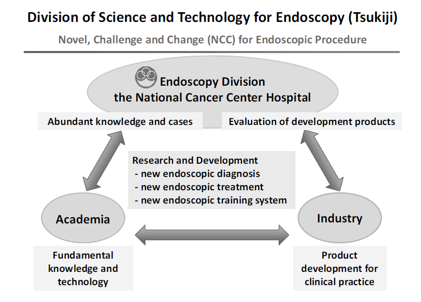
Since the birth of endoscope about half a century ago, endoscopes have undergone an era of fiber scopes. Now, with the introduction of videoscopes and high-definition systems, image quality has improved dramatically, and the target organs for observation and diagnosis have expanded from the stomach to include the esophagus, duodenum, colon, bronchus, biliary tract, and pharynx. The primary purpose of endoscopy is "observation and diagnosis" to detect lesions and check target organs; however, in recent years, "treatment" using endoscopes has become possible additionally. Nevertheless, currently, diagnosis is limited to focusing on morphological characteristics, and lesions that are missed occur at a certain rate, and difficult-to-treat lesions and treatment-related complications may also be encountered. In addition, the quality of endoscopic diagnosis and treatment often depends on the skill of endoscopists, and various issues remain to be resolved. To solve these issues and future demands, the Division of Science and Technology for Endoscopy, in collaboration with industry, government, and academia, is working to develop innovative endoscopic diagnosis, treatment, and training systems, and to disseminate them from Japan to the world (Figure 1).

Meanwhile, future demands include the development of innovative endoscopic diagnostic devices that visualize cancer characteristics, such as molecular imaging and functional imaging, and innovative endoscopic treatment techniques, such as endoscopic full-thickness resection. The Endoscopy Division of the National Cancer Center Hospital has a wealth of knowledge and clinical experience, which will play an effective role in the development of fundamental knowledge and technologies in academia, the development of products for actual clinical use and the evaluation and demonstration of developed products in industry.
The Team and What We Do
Activities began in fiscal year 2017, and in fiscal year 2023, each of the four teams (Diagnostic Team, Treatment Team, Training Team, and Endoscopic ultrasonography Team) continued to develop innovative endoscopic diagnosis, treatment, and training systems, with research results being gradually obtained.
Diagnosis Team: Naoya Toyoshima, Yasuhiko Mizuguchi, Haruhisa Suzuki, Satoru Nonaka, Masayoshi Yamada, Taku Sakamoto
Treatment Team: Satoru Nonaka, Seiichiro Abe, Hiroyuki Takamaru
Training Team: Haruhisa Suzuki, Nozomu Kobayashi, Yasuo Kadokawa, Naoya Toyoshima, Hiroyuki Takamaru, Keiko Nakamura
Endoscopic Ultrasonography Team: Susumi Hijioka, Shigetaka Yoshinaga
Supervisors: Shigetaka Yoshinaga, Yutaka Saito
At the Olympus Lab in the Corporate Collaboration Lab of the National Cancer Center Research Institute, we collaborate with Olympus Corporation to develop new endoscopic diagnostic, therapeutic, and training devices. The details are currently confidential, but each team is working on 3 to 4 themes.
Research Activities
1) Diagnosis Team
Length measuring device: The shape of 69 lesions was measured, and a formula was developed to predict the depth of lesions based on indices such as tension, which was evaluated in 53 lesions for a prospective study. In addition, spectroscopic measurements of lesions were performed to evaluate the optical spectral slope and change in lesion height.
Bio-optical phantoms: Under a joint research agreement between Olympus Corporation and NCCH, we measured lesions in the esophagus, stomach, and large intestine and accumulated data.
RDI Pit Pattern Color Difference Analysis: We have demonstrated that the combination of Red Dichromatic Imaging (RDI) and indigo carmine spraying facilitates the recognition of colorectal tumor lesion pit patterns by color difference analysis, and have been working on the publication of the results.
Improvement of quality of examination: To improve the quality of intragastric examination, we created a prototype model of the stomach with LEDs attached to the current model and developed an application to evaluate it.
Prevention of contamination of the lens of endoscopes: We investigated the prevention of contamination of the field of view of endoscopes from three perspectives: "evaluation of cleanability", "search for degradation factors" and "examination of cleaning techniques". We used pseudo-fouling models for "evaluation of cleanability," and "search for degradation factors. For the "examination of cleaning techniques," we searched for lens coating agents from both water repellent and hydrophilic viewpoints.
2) Treatment team
Suturing Devices: In collaboration with Olympus Corporation, we developed a stapler for soft endoscopes, and we conducted chronic animal experiments in August to confirm its clinical efficacy. However, after a value study within Olympus Corporation, they reached the conclusion that it was not profitable due to cost and the number of cases, and development at Olympus Corporation was terminated.
Platform for colonoscopy: We have been exploring the needs and cost estimates for a "device that is attached to the anus and supports endoscopic procedures," as well as an image of the device to reduce costs.
Effectiveness verification of large-diameter forceps holes: A brainstorming session was held in October, and we are reviewing the promotion system within Olympus.
3) Training Team
Endoscopic Operability: We studied the usefulness of endoscopic technique training using an endoscopy simplified training simulator, and demonstrated that this simulator is useful in training beginning physicians at the 103rd Annual Meeting of the Japanese Society of Gastrointestinal Endoscopy in May 2022. We then further explored new approaches using the endoscope simulator to simulate automatic operation by AI. Nevertheless, it is difficult for AI alone to complete the game autonomously, and we are now in the process of conducting a drastic review.
Colon insertion technology: In collaboration with Olympus, we are building a system to evaluate colonoscopy insertion techniques. As part of this review, we presented our efforts to quantify colonoscopy insertion operations using a training model at the 116th annual meeting of the Japan Gastroenterological Endoscopy Society, Kanto Chapter, in June 2023. In the future, as a new initiative, we plan to develop a new colonoscopy insertion training model based on the pain evaluation system developed by an academia and utilizing the know-how accumulated by Olympus and the three parties. We have conducted preliminary discussions with the academia.
Treatment Training Kit: For endoscopic treatment training, a pseudo gastrointestinal mucosa was created using konjak ingredients.
Education
Residents of the Endoscopy Division at the NCCH participate in the meetings of each development team and in various development experiments to train and educate the next generation of people who will be responsible for endoscopic device development.
Future Prospects
In the Division of Science and Technology for Endoscopy, we would like to work on the development of innovative endoscopic diagnosis, treatment, and training systems in collaboration with industry, academia, and government to solve many current issues and future demands in daily clinical practice, and to disseminate them from Japan to the world.
