Annual Report 2023
Department of Diagnostic Radiology
Tatsushi Kobayashi, Masayuki Yamaguchi, Hirofumi Kuno, Yasunori Arai, Kaoru Shimada, Tomoaki Sasaki, Takashi Hiyama, Shioto Oda, Takahiro Morita, Yohei Takei, Yusuke Miyasaka, Toshihiro Horii, Akihito Nakajima
Introduction
The Department of Diagnostic Radiology is committed to improving health through excellence in image-oriented patient care and research. Our department performs approximately 140,000 inpatient and outpatient examinations annually. Our department also conducts clinical scientific research and basic scientific studies, with the results translated directly into better patient care.
The Team and What We Do
Our department has four multi-slice CT scanners including one ultra-high-resolution CT, two area detector CT scanners and one dual-source CT, two 3T MRI systems, one interventional radiology (IR) CT system, one multi-axis c-arm CT system, two gamma cameras with the capacity for single photon emission CT (SPECT), two digital radiographic (DR) systems for fluoroscopy, two mammography (MMG) machines, and four computed radiographic (CR) systems. Our IR-CT systems use digital subtraction angiography with multi-detector computerized tomography (MDCT). One is equipped with a 320 multi-slice CT. A positron emission tomography (PET) scanner and baby cyclotron have been installed, and tumor imaging using 18F-FDG (fluorodeoxyglucose) has been performed. These all-digital image systems enhance the efficacy of routine examinations.
This department has eleven staff radiologists, a senior resident and a resident. As part of our routine activities, every effort is made to produce an integrated report covering almost all examinations, such as MMG, contrast radiological procedures, CT, MRI, RI, PET, angiography and IR, primarily transarterial chemoembolization (TACE).
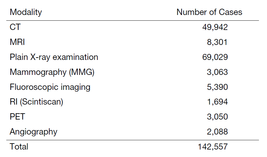
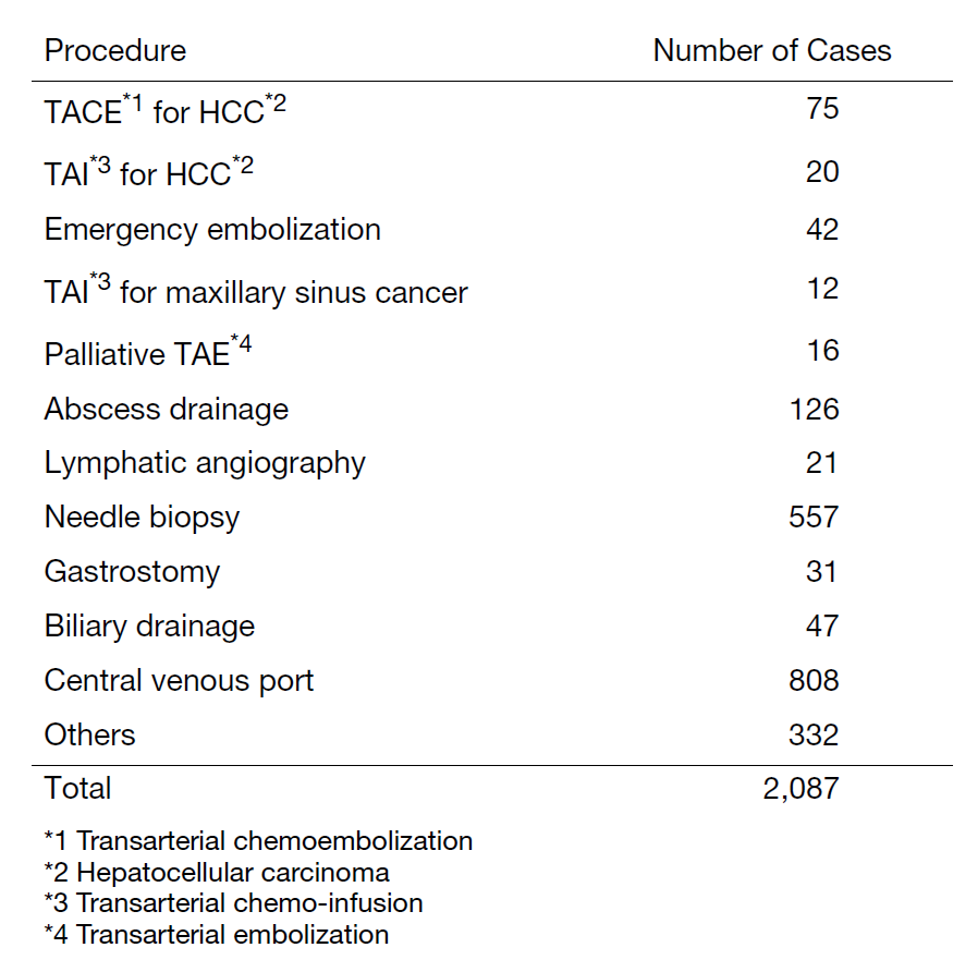
The number of cases examined in 2023 is shown in Table 1 and Table 2. Several conferences are routinely held at our department including pre- and postoperative conferences. Our department also contributes to deciding the treatment strategy through image presentations at the weekly tumor board conference.
Table 1. Number of examinations in 2023

Table 2. Number of Interventional RadiologyProcedures in 2023

Research Activities
The research activities of the Department of Diagnostic Radiology are focused on diagnostic imaging and IR. A major focus of our department is the development of new imaging techniques using advanced CT/MR systems, including ultra-high resolution CT (UHR CT), dual energy CT (DECT), and area detector CT (ADCT) for cancer patients. In addition, we have initiated clinical research with photon-counting CT, a technology that offers advantages such as higher spatial resolution and precise material differentiation through spectral imaging. Specifically, photon-counting CT offers several advantages: 1. Higher spatial resolution, 2. Superior signal-to-noise ratio allowing for lower radiation doses, 3. Improved image quality due to minimized electronic noise, 4. Precise material differentiation through spectral imaging. Spectral imaging and DECT technologies offer the potential to improve pathology detection and increase diagnostic confidence in the evaluation of a range of cancers. This is achieved by exploiting the x-ray energy-dependent absorption behavior of different materials. We are also exploring new image reconstruction techniques, in particular the AI-based Advanced Intelligent Clear-IQ Engine (AiCE). The overall goal is to improve diagnostic capabilities while reducing patient radiation exposure, thereby positioning the system as a valuable tool in clinical research. Our current work is primarily focused on lung and head and neck imaging, and we aim to compare these photon-counting CT systems with conventional CT platforms.
In addition to imaging technologies, our department is committed to developing new methods for making diagnoses and predicting prognosis, treatment response, and outcomes. We use radiomics and machine learning algorithms to accomplish this. One of our major efforts is to correlate tumor-specific radiomic features with clinical data, such as treatment outcomes. We are also researching the construction of image diagnosis technology that combines temporal subtraction technique and artificial intelligence. This technology is particularly useful for diagnosing cancer recurrence in patients. We are actively collaborating with various companies to develop and advance new AI technologies for imaging diagnosis.
Clinical Trials
We are conducting a multicenter clinical trial, "The single-armed confirmatory trial for immediate effectivity and safety of palliative arterial embolization for painful bone metastases. (JIVROSG/J-SUPPORT 1903)", as a representative facility, which was started in March 2021. The purpose of this trial is to verify the safety and immediate effect of transarterial embolization as a palliative treatment for painful bone metastases and to establish it as a standard treatment. This research is funded by the Japan Agency for Medical Research and Development (AMED).
List of papers published in 2023
Journal
1. Onodera K, Aokage K, Wakabayashi M, Ikeno T, Morita T, Ohashi S, Miyoshi T, Tane K, Samejima J, Tsuboi M. An accurate prediction of negative lymph node metastasis with consideration of glucose metabolism in early-stage non-small cell lung cancer. General thoracic and cardiovascular surgery, 72:24-30, 2024
2. Hiyama T, Miyasaka Y, Kuno H, Sekiya K, Sakashita S, Shinozaki T, Kobayashi T. Posttreatment Head and Neck Cancer Imaging: Anatomic Considerations Based on Cancer Subsites. Radiographics, 44:e230099, 2024
3. Komatsu M, Kitaguchi D, Yura M, Takeshita N, Yoshida M, Yamaguchi M, Kondo H, Kinoshita T, Ito M. Automatic surgical phase recognition-based skill assessment in laparoscopic distal gastrectomy using multicenter videos. Gastric cancer, 27:187-196, 2024
4. Ito K, Yamaguchi M, Semba T, Tabata K, Tamura M, Aoyama M, Abe T, Asano O, Terada Y, Funahashi Y, Fujii H. Amelioration of Tumor-promoting Microenvironment via Vascular Remodeling and CAF Suppression Using E7130: Biomarker Analysis by Multimodal Imaging Modalities. Molecular cancer therapeutics, 23:235-247, 2024
5. Sagara H, Inoue K, Yaku H, Ohsawa A, Mano C, Morita T, Hiyama T, Muramatsu Y, Inaki A, Fujii H. A new simpler image quality index based on body size for FDG-PET/CT. Nuclear medicine communications, 45:93-101, 2024
6. Nakama R, Inoue N, Miyamoto Y, Arai Y, Kobayashi T, Fushimi K. Patient characteristics and procedural and safety outcomes of percutaneous transesophageal gastro-tubing: A nationwide database study in Japan. Surgery, 175:368-372, 2024
7. Nakama R, Arai Y, Horii T, Kobayashi T. Computed tomography-guided percutaneous needle biopsy for middle mediastinal tumors with retroaortic paravertebral approach: A case report. Radiology case reports, 19:1440-1444, 2024
8. Sasaki T, Kuno H, Hiyama T, Oda S, Masuoka S, Miyasaka Y, Taki T, Nagasaki Y, Ohtani-Kim SJ, Ishii G, Kaku S, Shroff GS, Kobayashi T. 2021 WHO Classification of Lung Cancer: Molecular Biology Research and Radiologic-Pathologic Correlation. Radiographics, 44:e230136, 2024
9. Kunieda K, Makihara K, Yamada S, Yamaguchi M, Nakamura T, Terada Y. Brain Structures in a Human Embryo Imaged with MR Microscopy. Magnetic resonance in medical sciences, 2024
10. Miyasaka Y, Hiyama T, Kuno H, Shinozaki T, Sakashita S, Kobayashi T. Characteristic imaging findings in a patient with chronic expanding hematoma on the floor of the mouth. International cancer conference journal, 12:185-189, 2023
11. Kudo M, Gotohda N, Sugimoto M, Kobayashi S, Konishi M, Kobayashi T. Liver functional assessment using time-associated change in the liver-to-spleen signal intensity ratio on enhanced magnetic resonance imaging: a retrospective study. BMC surgery, 23:179, 2023
12. Oda S, Kuno H, Hiyama T, Sakashita S, Sasaki T, Kobayashi T. Computed tomography-based radiomic analysis for predicting pathological response and prognosis after neoadjuvant chemotherapy in patients with locally advanced esophageal cancer. Abdominal radiology (New York), 48:2503-2513, 2023
13. Masuoka S, Hiyama T, Kuno H, Sasaki T, Oda S, Miyasaka Y, Yamaguchi M, Kobayashi T. Computed tomography findings of hepatobiliary systems in patients with immune checkpoint inhibitor-induced liver injury. Abdominal radiology (New York), 48:3012-3021, 2023
14. Doan TKD, Umezawa M, Ikeda K, Ohnuki K, Akatsuka M, Okubo K, Kamimura M, Yamaguchi M, Fujii H, Soga K. Influence of Carboxyl Group Ratios on the Design of Breast Cancer Targeting Bimodal MR/NIR-II Imaging Probe from PLGA@Gd-DOTA@PEG Micelles Conjugating Herceptin. ACS applied bio materials, 6:2644-2650, 2023
15. Makihara K, Kunieda K, Yamada S, Yamaguchi M, Nakamura T, Terada Y. High-resolution MRI for human embryos with isotropic 10 µm resolution at 9.4 T. Journal of magnetic resonance (San Diego, Calif. : 1997), 355:107545, 2023
16. Hiyama T, Kuno H. Letter to Editor "Dual-energy CT for the detection of skull base invasion in nasopharyngeal carcinoma: comparison of simulated single-energy CT and MRI". Insights into imaging, 14:218, 2023
17. Oda S, Kuno H, Hiyama T, Sakashita S, Sasaki T, Kobayashi T. Reply to letter to the editor: Computed tomography-based radiomic analysis for predicting pathological response and prognosis after neoadjuvant chemotherapy in patients with locally advanced esophageal cancer. Abdominal radiology (New York), 48:3035-3037, 2023
