Annual Report 2023
Department of Radiological Technology
Masashi Ito, Masashi Shinozaki,Junpei Shigenaga, Yuichi Nagai, Aika Umemura, Kyohei Kuribayashi, Hiroki Miyazaki, Kaori Yanagisawa, Akira Inagaki, Moeka Mizuguchi, Kento Yokota, Honda Kazusa, Inoue Kanta, Kazuyuki Wakamatsu, Atsuko Takada, Naoki Yamazawa, Toshiyuki Shibuya, Shun Aoyagi, Hiroyuki Ohta, Mano Chikara, Takanori Matsuoka,Haruki Togo,Osawa Amon,Saeko Mochinaga,Manami Ihara,Hironori Kajiwara, Kotaro Takahashi, Harada Sumire, Aoki Yoshii, Tetsuya Aoyama, Yuki Miura, Risa Ohno, Suzuki Yuuta, Shinichi Takahashi, Mizunuma Yuki, Satoe Kito, Yuki Tanaka, Kazuhito Kano, Taku Tochiuchi, Hajime ohyoshi, Toshiya Rachi, Kota Hirotaki, Takashi Someya, Naoki Yamashita, Hashizume Nene, Ken Hirayama, Yuya Fujimoto, Yuuta Saito
Clinical Practice/Research Activities
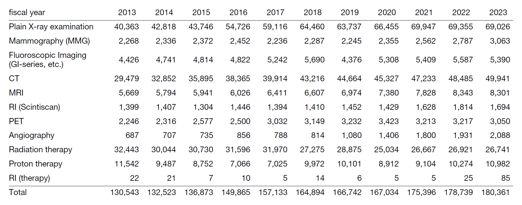
The total number of cases handled was 101% compared to the previous year, with no significant change (Table 1). In terms of details, in radiation therapy, the total number of proton beam therapy cases increased by 107%, but the number of patients remained almost unchanged. In radiological diagnosis, the number of CT examinations increased by 103%, as in previous years. In 2023, especially in RI treatment (internal therapy), a dedicated RI treatment ward was established and the number of cases increased by 340% compared to the previous year. It is expected that clinical trials for RI internal therapy will continue to shift from beta-emitting nuclides to alpha-emitting nuclides.
In research activities, we have signed joint research agreements with eight companies and have continued to conduct translational research on clinical technology, achieving results and even achieving commercialization. In addition, we are currently continuing our development of high-image-quality, high-performance photon-counting CT devices, and general radiography inspection systems using AI technology. In the field of radiation therapy, we are developing automatic plan check tools for radiation therapy plans, and are researching improvements and effective use of neutron activation suppressants in particle beam therapy facilities.
Table 1. Transitions in number of radiological examinations and radiation therapies by year.

Research Results
We conducted four years of collaborative research with a company using face recognition technology and developed an application that systemizes the entire process from reception to completion of CT examinations. We reported the results at the Radiological Society of North America (RSNA) and the European Congress of Radiology (ECR), and commercialized the application after confirming stable supply and quality maintenance.
We developed an automatic radiopharmaceutical management system for RI examinations that manages the amount of radiation used and the radiation exposure dose for patients based on information from the automatic radiopharmaceutical administration device. This research was also conducted in collaboration with a company for four years, and the research results were reported at the Radiological Society of North America and the European Congress of Radiology, and the product was commercialized.
In X-ray mammography examinations, we conducted four years of collaborative research with a company on "production of a deformable breast phantom for X-ray mammography accuracy management," and commercialized the product. The phantom, a model similar to a clinical breast, is a product that is expected to be used for automatic detection of lesions due to breast compression.
In the field of radiation therapy, in head and neck radiation therapy, more stable position matching has become possible by analyzing positional errors of anatomical structures that affect dose distribution. We also developed a fully automated program for intensity-modulated radiation therapy planning for locally advanced rectal cancer.
Education
Job rotation is carried out within the hospital to maintain and improve the functions of each department. In the Department of Radiological Technology, seven staff members work in each department, including the Clinical Coordinator's Office, Department of Medical Information, Ethical Review Support Section, Medical Affairs Division, Department for the Promotion of Medical Device Innovation, and seconded to the Japan Agency for Medical Research and Development (AMED). In addition, the radiological technologist resident system was launched in April 2022, and the first, second, and third classes (1, 2, and 2, respectively) are currently in progress according to the syllabus and curriculum. The purpose of this system is to train and produce diagnostic radiological technologists with highly specialized knowledge and skills to equalize, develop, and expand cancer care, and to protect the lives and health of the people. This system consists of two stages: a three-year basic course for cancer specialists (general medical training course at the Kashiwa campus) and a two-year cancer specialist training course. 26 people participated in the online information session for the 2025 resident recruitment, and 7 people attended the tour.
Future Prospects
As the workload continues to increase, and in light of the shift in tasks, the hospital aims to provide training for the expansion of the scope of work and increase the number of people with various certifications. In order to carry out high-quality medical care, research, and education, the current staff is expected to achieve more visible results and accomplishments.
