Annual Report 2024
Department of Diagnostic Radiology
Tatsushi Kobayashi, Masayuki Yamaguchi, Hirofumi Kuno, Yasunori Arai, Kaoru Shimada, Tomoaki Sasaki, Takashi Hiyama, Shioto Oda, Takahiro Morita, Yohei Takei, Yusuke Miyasaka, Toshihiro Horii, Akihito Nakajima.
Introduction
The Department of Diagnostic Radiology is committed to advancing healthcare through excellence in image-guided patient care and research. Our department performs approximately 140,000 inpatient and outpatient examinations annually. We also pursue a comprehensive program of clinical and basic science research, directly translating our findings into improved patient care outcomes.
The Team and What We Do
Our department operates four multi-detector CT scanners: one ultra-high-resolution CT, two area-detector CT scanners, and one dual-source CT. We also have two 3T MRI systems, one interventional radiology (IR) CT system, one multi-axis C-arm CT system, two gamma cameras capable of SPECT, two digital radiographic (DR) fluoroscopy systems, two mammography units, and four computed radiography (CR) systems. Our IR-CT systems use digital subtraction angiography with 320-row multi-detector CT. A positron emission tomography (PET) scanner and baby cyclotron have been installed, and tumor imaging using 18F-FDG (fluorodeoxyglucose) has been performed. These all-digital image systems enhance the efficacy of routine examinations.
This department has eleven staff radiologists, a senior resident and a resident. As part of our routine activities, every effort is made to produce an integrated report covering almost all examinations, such as MMG, contrast radiological procedures, CT, MRI, RI, PET, angiography and IR, primarily transarterial chemoembolization (TACE).
The department plays a vital role in patient management by actively participating in numerous preoperative and postoperative conferences. Furthermore, our imaging presentations at the weekly cancer board meetings are crucial in determining optimal treatment strategies for patients. The case volumes for 2024 are detailed in Tables 1 and 2.
Table 1. Number of examinations in 2024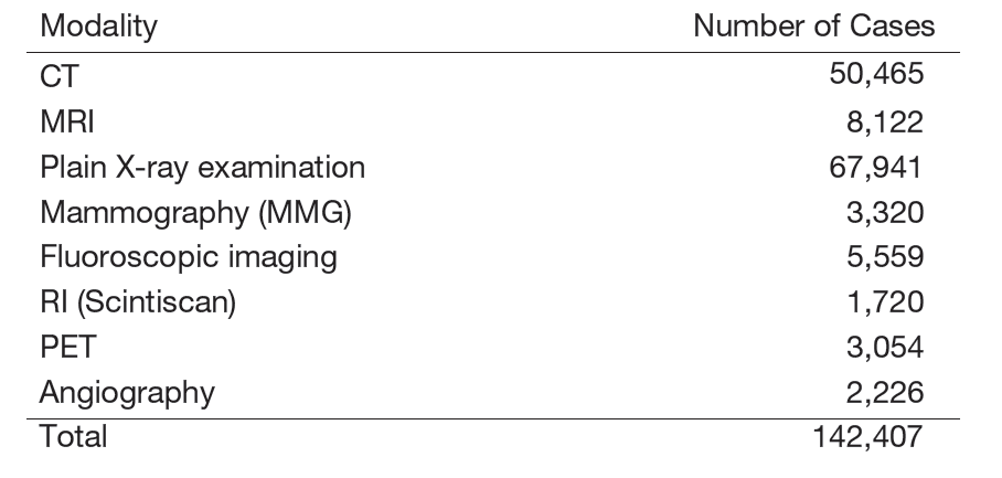
Table 2. Number of Interventional Radiology Procedures in 2024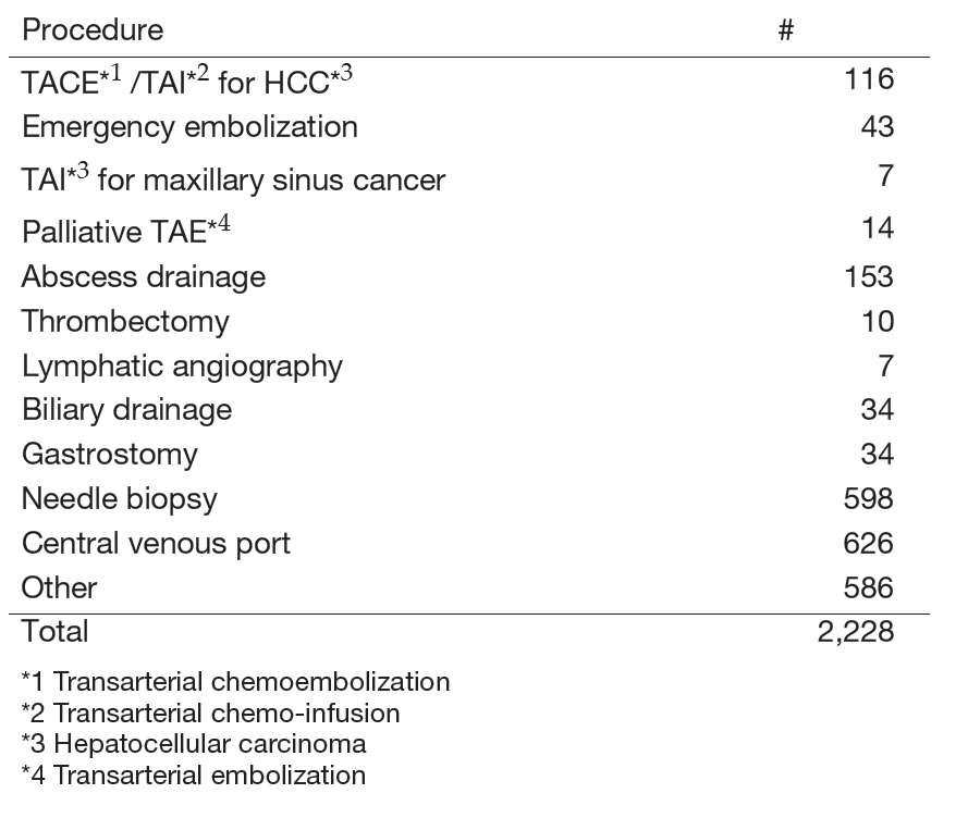
Research Activities
Our research activities focus on advanced diagnostic imaging and interventional radiology, developing innovative imaging techniques using advanced CT and MR systems, including ultra-high-resolution CT (UHR-CT), dual-energy CT (DECT), and area detector CT (ADCT) for oncological applications. We have initiated clinical research with photon-counting CT, which offers multiple advantages: enhanced spatial resolution, superior signal-to-noise ratios, reduced radiation dose, improved image quality through minimized electronic noise, and precise material differentiation via spectral imaging. Photon-counting CT and spectral imaging technologies can improve pathological detection and increase diagnostic confidence across a range of cancers. These technologies exploit multi-energy X-ray data acquisition for precise material decomposition analysis. We are investigating novel spectral reconstruction techniques, particularly low-energy virtual monoenergetic imaging at 40keV which significantly enhances contrast resolution. Our overarching goal is to enhance diagnostic capabilities while minimizing patient radiation exposure. Our current research focuses on lung, head and neck, and pancreatic imaging, as well as comparative studies between photon-counting CT systems and conventional CT platforms.
We also develop innovative methods for diagnosis, prognosis prediction, treatment response assessment, and outcome evaluation using radiomics and deep learning algorithms. We research imaging diagnostic technology that combines temporal subtraction techniques with artificial intelligence, which is particularly valuable for diagnosing cancer recurrence. We collaborate with various industry partners to develop and advance new AI technologies for imaging diagnosis.
Clinical Trials
We are conducting a multicenter clinical trial, "The single-armed confirmatory trial for immediate effectivity and safety of palliative arterial embolization for painful bone metastases. (JIVROSG/J-SUPPORT 1903)", as a representative facility, which was started in March 2021. The purpose of this trial is to verify the safety and immediate effect of transarterial embolization as a palliative treatment for painful bone metastases and to establish it as a standard treatment. This research is funded by the Japan Agency for Medical Research and Development (AMED).
List of papers published in 2024
Journal
1. Sakai M, Hiyama T, Kuno H, Kobayashi T, Nakajima T. Imaging of the skull base and orbital tumors. Japanese journal of radiology, 43:152-163, 2025
2. Oda S, Kuno H, Fujita T, Hiyama T, Kotani D, Kadota T, Sakashita S, Kobayashi T. Clinical usefulness of four-dimensional dynamic ventilation CT for borderline resectable locally advanced esophageal cancer. Japanese journal of radiology, 43:434-444, 2025
3. Sasaki T, Oda S, Kuno H, Hiyama T, Taki T, Takahashi S, Ishii G, Tsuboi M, Kobayashi T. Potential of spectral imaging generated by contrast-enhanced dual-energy CT for lung cancer histopathological classification - A preliminary study. European journal of radiology open, 14:100628, 2025
4. Ito K, Kuno H, Otsuka K, Andreu-Arasa VC, Sakai O, Kaneda T. Imaging Findings, Complications, and Mimics after Common and Advanced Dental Procedures. Radiographics : a review publication of the Radiological Society of North America, Inc, 45:e240072, 2025
5. Sasaki T, Kuno H, Nomura K, Muramatsu Y, Aokage K, Samejima J, Taki T, Goto E, Wakabayashi M, Furuya H, Taguchi H, Kobayashi T. CZT-based photon-counting-detector CT with deep-learning reconstruction: image quality and diagnostic confidence for lung tumor assessment. Japanese journal of radiology, 2025
6. Sasaki T. Long-Term Prognostic Implications of Thoracic Aortic Calcification and Coronary Artery Calcification on Low-Dose CT in a Screening Population From East Asia. AJR. American journal of roentgenology, 2025
7. Miyasaka Y, Hiyama T, Kuno H, Shinozaki T, Tomioka T, Sakashita S, Kobayashi T. Imaging of salivary gland cancers derived from a sublingual gland herniated into the submandibular space: a report of three cases. Neuroradiology, 66:931-935, 2024
8. Oda S, Kuno H, Hiyama T, Yamaguchi M, Sasaki T, Yajima S, Masuda H, Kobayashi T. Radiologic feature of complications after artificial urinary sphincter implantation following total prostatectomy. Abdominal radiology (New York), 49:2416-2427, 2024
9. Henson C, Abou-Foul AK, Yu E, Glastonbury C, Huang SH, King AD, Lydiatt WM, McDowell L, Nagelschneider AA, Nankivell PC, O'Sullivan B, Rhys R, Xiao Y, Andrew D, Asmussen JT, Bidault F, Dankbaar JW, de Graaf P, Gebrim ES, Hu C, Ding J, Kanda T, Kim J, Kuno H, Medrano-Martorell S, Oikonomopoulos N, Goh JP, Santos-Armentia E, Schafigh DG, Subramaniam RM, Wu XC, Yom SS, Mehanna H. Criteria for the diagnosis of extranodal extension detected on radiological imaging in head and neck cancer: Head and Neck Cancer International Group consensus recommendations. The Lancet. Oncology, 25:e297-e307, 2024