Home > Organization > Division of Advanced Bioimaging
Division of Advanced Bioimaging

- Feb. 26, 2026
- Mr. Toshiki Mori received the Director's Award at the 10th Tsukiji Campus Young Staff Research Presentation Meeting.
- Dec. 18, 2025
- Kenichi Suzuki gave a presentation at Pacifichem 2025 (Honolulu, USA).
- Dec. 16, 2025
- Kenichi Suzuki gave a presentation at Pacifichem 2025 (Honolulu, USA).
- Nov. 12, 2025
- Kenichi Suzuki gave a keynote lecture at the 2025 Annual Meeting of the Society for Glycobiology (San Diego, USA).
- Aug. 14, 2025
- The main paper in our research field has been published in iScience
- Jul. 10, 2025
- Kenichi Suzuki gave a talk at Biophysics society Thematic Meeting (Copenhagen, Denmark).
- Jul. 1, 2025
- Koichiro Hirosawa joined our lab as a specially appointed researcher.
- Jun. 4, 2025
- Kenichi Suzuki gave a special lecture at the 47th Annual Meeting of the Membrane society of Japan
- Apr. 30, 2025
- The main paper in our research field has been published in Journal of Cell Biology
- Mar. 27, 2025
- Kenichi Suzuki delivered a talk at Gordon Research Conference (Lucca, Italy)

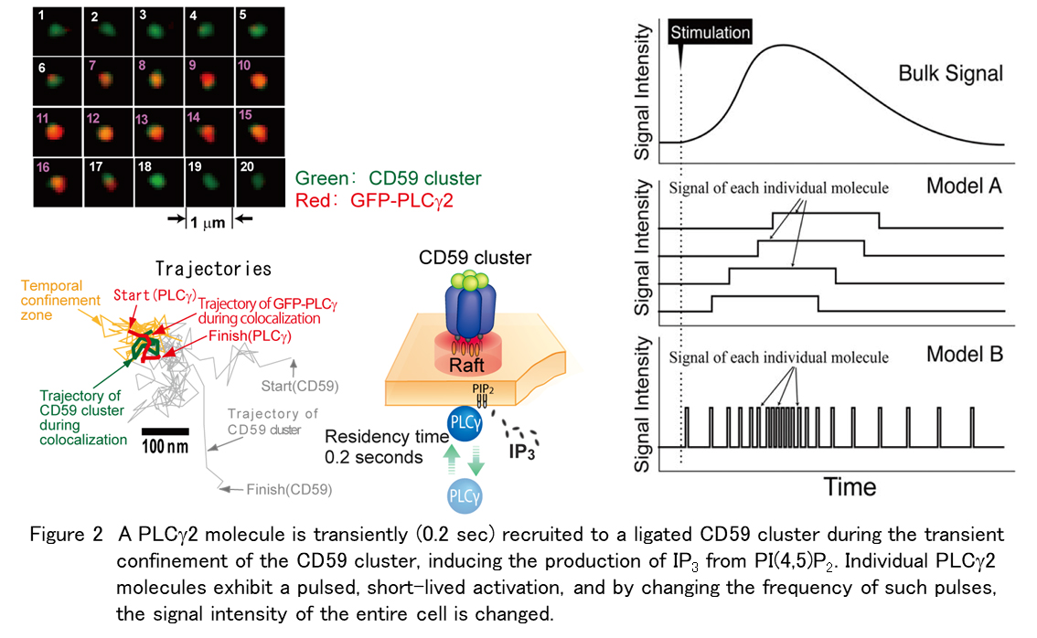
Molecules in a cell do not work synchronously but essentially work in a stochastic process theory (randomly). Individual molecules engage in interactions lasting mere seconds, with the proportion of interacting molecules rarely exceeding 10%. For example, single-molecule observation revealed that an oncogene product, K-Ras is activated by binding with GTP for only less than 0.5 seconds, and the activated fraction is less than 10% (Figure 1). We have found that changes in the bulk signal of the entire cell, such as phosphorylation of signaling molecules observed by western blotting and other methods, usually last from several to 20 minutes, but that each molecule causes a pulsed, short-lived signal, and by changing the frequency of such pulses, the signal intensity of the entire cell is changed (Suzuki et al., J. Cell Biol., 2007a,b; Figure 2).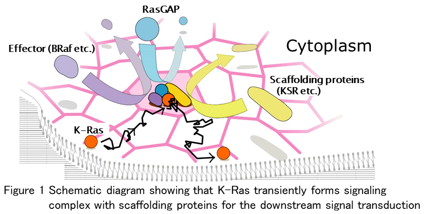
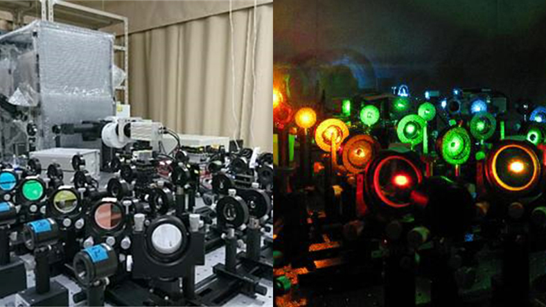
Therefore, in order to elucidate the molecular mechanisms in cells, we believe that the best way is to observe individual molecules in cells (sometimes at the world’s fastest speed of 10,000 frames per second!) and collect statistics on the time and frequency of events. (Naturally, the results obtained via single-molecule imaging must correspond with those obtained through observations of multiple molecules) We hope to elucidate from the first principles the signaling and regulatory mechanisms associated with cellular oncogenesis by single-molecule and super-resolution microscopy and use this information for clinical and drug discovery purposes.